
Session Information
Date: Wednesday, October 24, 2018
Title: 6W011 ACR Abstract: Spondyloarthritis Incl PsA–Clinical VI: Imaging of Axial SpA (2922–2927)
Session Type: ACR Concurrent Abstract Session
Session Time: 9:00AM-10:30AM
Background/Purpose: Fatty lesions (FL), similar to bone marrow edema (BME) and sclerosis (SCL), are characteristic findings in MRI examinations of patients with ankylosing spondylitis (AS) and degenerative disc disease (DDD). It has recently been shown that FL are associated with syndesmophyte formation in AS. The anatomic correlate of FL has not been studied to date. Current assumptions are solely based on non-invasive data. Here we examine the cellular composition of FL in the edges of vertebral bodies of patients with AS or DDD by histology.
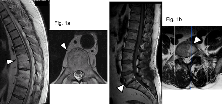
Methods: Patients with AS or DDD undergoing planned kyphosis correction surgery by spinal osteotomy (in AS) or surgery to correct spinal stenosis (in DDD) were included into this biopsy study. The spinal surgeon (HB) took all biopsies mainly in the area close to the vertebral edge in many of which FL had been seen by MRI (Fig. 1a for AS and 1b for DDD). Biopsies were decalcified, embedded in paraffin, cut and stained by hematoxylin and eosin. The marrow composition was analyzed and the cellularity graded (% surface area) by two different investigators blinded to patients’ diagnosis. Four different marrow compositions could be differentiated: (i) fat, (ii) fibrosis, (iii) inflammation and (iv) hematopoiesis (normal).
Results: A total of 60 biopsies mostly obtained from the lower thoracic spine and the lumbar spine of 21 AS patients (mean age 51.7 years, mean disease duration 24.6 years) and of the lumbar spine in 18 DDD patients (mean age 60.1 years) were available. On the patient level, the histological appearance of MRI-FL was different between the groups: fat marrow was present in biopsies of 19 AS (90%) but in only 5 DDD (28%) patients. Inflammatory marrow changes, resembling mononuclear infiltrates, were found in 8 AS (38.1%) and 14 DDD (77.8%) patients at areas with concomitant FL and BME on MRI, while marrow fibrosis was seen in 6 AS (28.6%) and 4 DDD (22.2%) patients at areas with concomitant FL and SCL on MRI.
In the semiquantitative histopathological analysis, the mean distribution (±standard deviation) of the various bone marrow tissue types in the biopsies differed between the AS vs. DD in a similar way, with 43% (±26.3%) vs. 16% (±30.3%) for fatty marrow, 11% (±15.5%) vs. 55% (±42%) for inflammatory marrow and 9% (±16.1%) vs. 13% (±27.8%) for fibrotic marrow, respectively.
Conclusion: The presence of FL on MRI corresponds to fat deposition in the bone marrow of patients with advanced AS. These data show that the MRI change termed “fatty lesion” is indeed based on the deposition of fat in the vertebral bone marrow in AS. Since vertebral bone marrow is physiologically harboring hematopoiesis, AS seems to lead to a change in the bone marrow microenvironment with local disruption of hematopoiesis and replacement by fat. The link between fat and new bone formation should be studied in earlier disease stages.
To cite this abstract in AMA style:
Baraliakos X, Boehm H, Samir A, Schett G, Braun J, Ramming A. Fatty Lesions Detected on MRI Scans in Patients with Ankylosing Spondylitis Are Based on the Deposition of Fat in the Vertebral Bone Marrow [abstract]. Arthritis Rheumatol. 2018; 70 (suppl 9). https://acrabstracts.org/abstract/fatty-lesions-detected-on-mri-scans-in-patients-with-ankylosing-spondylitis-are-based-on-the-deposition-of-fat-in-the-vertebral-bone-marrow/. Accessed .« Back to 2018 ACR/ARHP Annual Meeting
ACR Meeting Abstracts - https://acrabstracts.org/abstract/fatty-lesions-detected-on-mri-scans-in-patients-with-ankylosing-spondylitis-are-based-on-the-deposition-of-fat-in-the-vertebral-bone-marrow/