Session Information
Session Type: Abstract Submissions (ACR)
Background/Purpose: Altered tibiofemoral (TF) joint kinematics and joint surface interactions have been linked with development and progression of knee osteoarthritis (OA). However, accurate in-vivo investigations of the TF joint mechanics in patients with knee OA have been difficult to perform due to limitations of conventional techniques. This study sought to accurately characterize TF joint kinematics and the interactions of the articulating joint surfaces using high-speed dynamic stereo x-ray (DSX) technology during the loading phase of downhill walking in older adults with and without knee OA.
Methods: Eleven subjects with knee OA and 10 subjects without OA participated in this study. Subjects with knee OA were included if they demonstrated a Kellgren and Lawrence radiographic OA severity of at least grade II or higher. High speed DSX images were acquired during the loading phase of a moderately declined walking condition (7% grade, 0.75 m/s) on an instrumented treadmill. Computerized tomography images were also taken to create subject-specific 3D bone models of the distal femur and proximal tibia. A previously validated model-based tracking algorithm was employed to determine 3D joint motion by matching the radiographic images with projections through the volumetric bone models. The anterior/posterior (AP) and medial/lateral (ML) positions of the TF joint contact points were estimated using the distance-weighted centroids of the region of closest bony proximity in both the medial and lateral TF compartments. ML and AP contact path lengths were determined by subtracting the minimum from the maximum AP and ML contact positions. In addition, the total contact path was determined as the algebraic summation of the ML and AP translations of the contact points in each compartment.
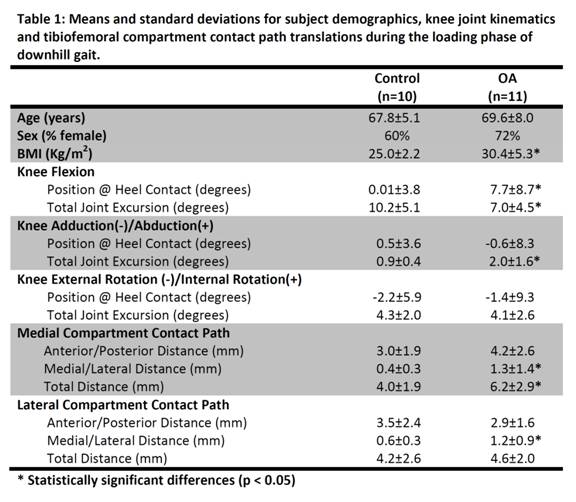
Results: Compared to the control group, subjects with knee OA contacted the ground with more knee flexion but moved through less flexion range of motion. Conversely, subjects with knee OA moved through more knee adduction range of motion despite contacting the ground with a near neutral frontal plane knee alignment. Additionally, the OA group demonstrated longer ML and total contact path lengths for the medial TF compartment and a longer ML contact path for the lateral TF compartment.
Conclusion: Consistent with previous reports, subjects with knee OA contacted the ground with more knee flexion. However, findings from this study further suggest that individuals with knee OA also move through less knee flexion range of motion which can adversely affect shock absorption. Additionally, individuals with knee OA demonstrated signs of frontal plane TF joint instability (excessive adduction motion and ML translation) and a longer medial TF compartment translation which can negatively impact the rate of OA progression. Intervention strategies to improve knee flexion during loading while limiting excessive frontal plane motion and joint translations should be considered.
Disclosure:
S. Farrokhi,
None;
C. A. Rainis,
None;
G. K. Fitzgerald,
None;
S. Tashman,
None.
« Back to 2012 ACR/ARHP Annual Meeting
ACR Meeting Abstracts - https://acrabstracts.org/abstract/dynamic-stereo-x-ray-evaluation-of-knee-joint-mechanics-during-downhill-walking-in-subject-with-knee-osteoarthritis/

