ACR Meeting Abstracts

ACR Meeting Abstracts

  • Meetings
    • ACR Convergence 2024
    • ACR Convergence 2023
    • 2023 ACR/ARP PRSYM
    • ACR Convergence 2022
    • ACR Convergence 2021
    • ACR Convergence 2020
    • 2020 ACR/ARP PRSYM
    • 2019 ACR/ARP Annual Meeting
    • 2018-2009 Meetings
    • Download Abstracts
  • Keyword Index
  • Advanced Search
  • Your Favorites
    • Favorites
    • Login
    • View and print all favorites
    • Clear all your favorites
  • ACR Meetings

Abstract Number: 0037

Dual (and Dueling) Fibroblast Differentiation Is Integral to the Pathogenesis of Anti-Ro Mediated Congenital Heart Block and Provides a Novel Biomarker of Severity

Christina Firl1, Robert Clancy2, Bettina Cuneo3, Nicola Fraser4, Marc Halushka5, Mala Masson6, Amit Saxena1 and Jill Buyon2, 1NYU Langone Health, New York, NY, 2NYU Grossman School of Medicine, New York, NY, 3University of Colorado School of Medicine, Aurora, CO, 4NYU School of Medicine, New York, NY, 5Johns Hopkins Medicine, Baltimore, MD, 6NYU Langone Medical Center- Division of Rheumatology, New York, NY

Meeting: ACR Convergence 2022

Keywords: Autoantibody(ies), Fibroblasts, Other, Fibrosing syndromes

  • Tweet
  • Click to email a link to a friend (Opens in new window) Email
  • Click to print (Opens in new window) Print
Session Information

Date: Saturday, November 12, 2022

Title: Pediatric Rheumatology – Basic Science Poster

Session Type: Poster Session A

Session Time: 1:00PM-3:00PM

Background/Purpose: Fibroblast heterogeneity may provide critical insights into autoimmune mediated tissue damage. Transdifferentiation to a scarring myofibroblast is characterized by expression of alpha-Smooth Muscle Actin (α-SMA), deposition of collagen and contractile properties while another fibroblast subtype evaluated largely in tumor migration is distinguished by expression of S100 Calcium Binding Protein A4 (S100A4). Given the histologic hallmark of AV nodal fibrosis and endocardial fibroelastosis in anti-Ro mediated cardiac neonatal lupus (NL), this study addressed the contribution of S100A4 and α-SMA-expressing fibroblasts.

Methods: In an in vitro model of cardiac-NL, 2nd trimester human fetal cardiac fibroblasts were co-cultured with supernatants (sup) of macrophage-like THP-1 cells transfected with hY3 (noncoding ssRNA TLR7/8 agonist binding Ro60) and additional treatments with readouts: immunofluorescence, flow cytometry, qPCR, and a collagen assay. S100A4 was measured in anti-Ro exposed cord bloods and expression evaluated in fetal cardiac tissue.

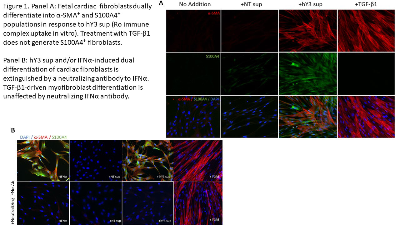
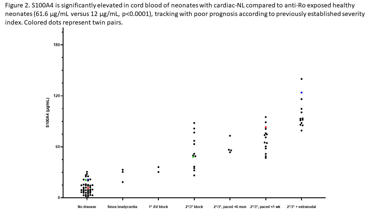
Results: When cultured with hY3 sup, cardiac fibroblasts differentiated into two populations, distinguished by expression of α-SMA or S100A4. In contrast, activated TGF-β1 only resulted in expression of α-SMA (Figure 1). These results were confirmed by flow cytometry; hY3 sup promoted expression of both α-SMA (8263±5185) and S100A4 (1316±469) whereas TGF-β1 stimulated fibroblasts expressed α-SMA (9085±5867) not S100A4 (159.2±56.9) (p=0.002, N=4). Unlike TGF-β1 but similar to hY3 sup, IFNα induced dual fibroblast differentiation (α-SMA-16332±9085, S100A4-1698±806.4, p=0.008, N=3). Dual differentiation induced by hY3 sup or IFNα was inhibited by anti-IFNα which had no effect on TGF-β1-induced α-SMA. hY3-treated fibroblasts upregulated transcription of proangiogenic cytokines IL6, IL10 and MMP13 (ECM remodeling) compared to TGF-β1 (5.7 vs 1.5, p=0.0079, N=5; 5.1 vs 0.96, p=0.032, N=3; 7.5 vs 0.8, p=0.0079, N=5) while expression indicative of collagen deposition (Col1α1) was upregulated by TGF-β1 compared to hY3 sup (4.8 vs 1.8, p=0.028, N=4). In distinction, S100A4-positive fibroblasts uniquely degraded soluble collagen (14 ug/mL to 0.6, p=0.026, N=5). Bolstering in vivo relevance, circulating S100A4 was elevated in cardiac-NL (N=49) compared to non-cardiac-NL (N=47) (61.6 vs 12.0 ug/mL, p< 0.0001). Remarkably, levels tracked with an established cardiac severity score and in twins, distinguished the affected from the unaffected neonate (Figure 2). S100A4 immunostaining was observed in 3 cardiac-NL hearts focally in fibroblasts within the conducting tissue but not in a healthy heart. α-SMA was detected in fibrotic zones while S100A4 in regions with dystrophic calcification.

Conclusion: In vitro and in vivo experiments position the S100A4-positive fibroblast subtype alongside the canonical myofibroblast in the pathogenesis of cardiac-NL. The pro-angiogenic and distinct collagenase activity may represent a thwarted attempt to mitigate fibrotic sequelae by the collagen-secreting myofibroblast with resultant unintended soft tissue calcification. Moreover neonatal S100A4 levels support a novel biomarker of poor prognosis.

Supporting image 1

Supporting image 2


Disclosures: C. Firl, None; R. Clancy, None; B. Cuneo, None; N. Fraser, None; M. Halushka, None; M. Masson, None; A. Saxena, None; J. Buyon, Equillium, GlaxoSmithKlein(GSK), L and M Healthcare Communications, Janssen, Boomcom, Merck/MSD.

To cite this abstract in AMA style:

Firl C, Clancy R, Cuneo B, Fraser N, Halushka M, Masson M, Saxena A, Buyon J. Dual (and Dueling) Fibroblast Differentiation Is Integral to the Pathogenesis of Anti-Ro Mediated Congenital Heart Block and Provides a Novel Biomarker of Severity [abstract]. Arthritis Rheumatol. 2022; 74 (suppl 9). https://acrabstracts.org/abstract/dual-and-dueling-fibroblast-differentiation-is-integral-to-the-pathogenesis-of-anti-ro-mediated-congenital-heart-block-and-provides-a-novel-biomarker-of-severity/. Accessed .
  • Tweet
  • Click to email a link to a friend (Opens in new window) Email
  • Click to print (Opens in new window) Print

« Back to ACR Convergence 2022

ACR Meeting Abstracts - https://acrabstracts.org/abstract/dual-and-dueling-fibroblast-differentiation-is-integral-to-the-pathogenesis-of-anti-ro-mediated-congenital-heart-block-and-provides-a-novel-biomarker-of-severity/

Advanced Search

Your Favorites

You can save and print a list of your favorite abstracts during your browser session by clicking the “Favorite” button at the bottom of any abstract. View your favorites »

All abstracts accepted to ACR Convergence are under media embargo once the ACR has notified presenters of their abstract’s acceptance. They may be presented at other meetings or published as manuscripts after this time but should not be discussed in non-scholarly venues or outlets. The following embargo policies are strictly enforced by the ACR.

Accepted abstracts are made available to the public online in advance of the meeting and are published in a special online supplement of our scientific journal, Arthritis & Rheumatology. Information contained in those abstracts may not be released until the abstracts appear online. In an exception to the media embargo, academic institutions, private organizations, and companies with products whose value may be influenced by information contained in an abstract may issue a press release to coincide with the availability of an ACR abstract on the ACR website. However, the ACR continues to require that information that goes beyond that contained in the abstract (e.g., discussion of the abstract done as part of editorial news coverage) is under media embargo until 10:00 AM ET on November 14, 2024. Journalists with access to embargoed information cannot release articles or editorial news coverage before this time. Editorial news coverage is considered original articles/videos developed by employed journalists to report facts, commentary, and subject matter expert quotes in a narrative form using a variety of sources (e.g., research, announcements, press releases, events, etc.).

Violation of this policy may result in the abstract being withdrawn from the meeting and other measures deemed appropriate. Authors are responsible for notifying colleagues, institutions, communications firms, and all other stakeholders related to the development or promotion of the abstract about this policy. If you have questions about the ACR abstract embargo policy, please contact ACR abstracts staff at [email protected].

Wiley

  • Online Journal
  • Privacy Policy
  • Permissions Policies
  • Cookie Preferences

© Copyright 2025 American College of Rheumatology