Session Information
Session Type: Abstract Submissions (ACR)
Background/Purpose
It is assumed that persons with hip pain from osteoarthritis (OA) are likely to have radiographic OA, making it possible to readily diagnose disease, but there is little data addressing the agreement of hip pain with x-ray OA. We previously reported poor agreement between hip pain and RHOA (radiographic hip osteoarthritis) in the Framingham population. However, the Framingham study used long limb x-rays that may have yielded imperfect RHOA estimates. We examined concordance of hip pain and RHOA in the Osteoarthritis Initiative (OAI) where subjects obtained pelvis x-rays and were asked a more comprehensive set of questions about hip pain.
Methods
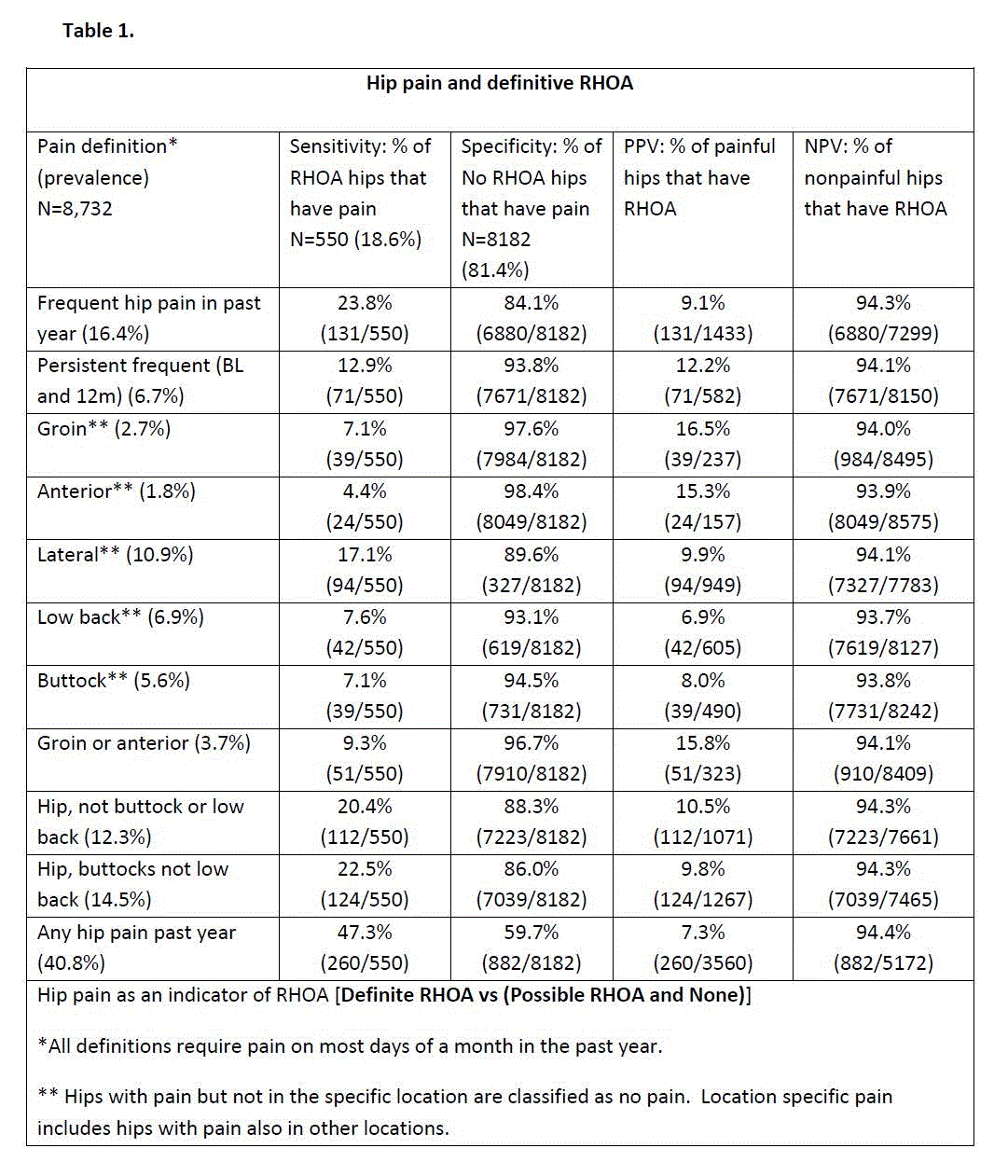
OAI is a multicenter longitudinal cohort study of OA that included 4796 individuals aged 45-79. AP pelvis x-rays were obtained, and definite RHOA was defined using UCSF criteria: 1) definite osteophytes plus definite JSN (both score ≥ 2) OR 2) definite osteophytes or definite JSN plus sclerosis, cysts or femoral head flattening OR 3) definite femoral osteophytes regardless of other features OR 4) definite moderate-severe JSN (superolateral JSN >=2 or superomedial JSN >=3) regardless of other features. Using a card with visual homunculus, subjects were asked whether they had hip pain on most days in a month. Those who said ‘yes’ were defined as having frequent hip pain and were asked another question for location of pain: groin, front of the leg (anterior), outside the leg (lateral), lower back, buttocks, or ‘don’t know’. The pain evaluation was done for both hips. We examined sensitivity (Sn), specificity (Sp) and positive and negative predictive values (PPV, NPV) for location specific pain with RHOA. Sn was defined as % of hips with RHOA that had hip pain. PPV was % of hips with pain that have RHOA. To ensure that we included hips that may have OA and to increase our sensitivity, we did another analysis for possible RHOA.
Results
X-rays from 8732 hips were evaluated. The prevalence of definite RHOA was 6.3%, and possible RHOA was 12.3%. For definite RHOA, the Sn of frequent hip pain was only 23.8%, and the Sp was 84.1% and the PPV for hip pain was only 9.1% (table 1). However, for analysis restricted to hip pain localized to the groin, the PPV rose to 16.5%. Of those with RHOA, only 7.1% had pain localized to the groin. Anterior hip pain resulted similarly to groin pain, but performance for other sites was diagnostically poorer. For possible RHOA (data not shown), the diagnostic test performance did not differ greatly from definite RHOA.
Conclusion
We found poor agreement between hip pain on most days and RHOA in the ipsilateral hip. Hip pain questions with the highest PPV were hip pain with groin or anterior pain, but most persons with this pain had negative radiographs for hip OA suggesting that x-rays are insensitive to the presence of disease. Many middle aged and older persons with chronic hip joint area pain may have OA even though x-rays are negative.
Disclosure:
C. Kim,
None;
M. C. Nevitt,
None;
P. M. Jungmann,
None;
I. Tolstykh,
None;
N. E. Lane,
None;
T. M. Link,
None;
D. T. Felson,
None.
« Back to 2014 ACR/ARHP Annual Meeting
ACR Meeting Abstracts - https://acrabstracts.org/abstract/discordance-of-hip-pain-with-radiographic-hip-osteoarthritis-the-osteoarthritis-initiative/

