ACR Meeting Abstracts

ACR Meeting Abstracts

  • Meetings
    • ACR Convergence 2024
    • ACR Convergence 2023
    • 2023 ACR/ARP PRSYM
    • ACR Convergence 2022
    • ACR Convergence 2021
    • ACR Convergence 2020
    • 2020 ACR/ARP PRSYM
    • 2019 ACR/ARP Annual Meeting
    • 2018-2009 Meetings
    • Download Abstracts
  • Keyword Index
  • Advanced Search
  • Your Favorites
    • Favorites
    • Login
    • View and print all favorites
    • Clear all your favorites
  • ACR Meetings

Abstract Number: 1617

Direct Comparison of Ultrasound, [18F]Fluorodeoxyglucose-Positron Emission Tomography/Computed Tomography and Magnetic Resonance Imaging: Early Diagnostics in Patients Suspected of Giant Cell Arteritis

Marieke van Nieuwland1, Marloes Vermeer1, Edgar Colin2, Nils Wagenaar1, Onno Vijlbrief1, Jordy van Zandwijk3, Riemer Slart4, Hendrik Koffijberg3, Erik Groot Jebbink3, Elisabeth Brouwer4, Dennis Boumans1 and Celina Alves1, 1Hospital Group Twente, Almelo, Netherlands, 2Hospital Group Twente, Deventer, Netherlands, 3University of Twente, Enschede, Netherlands, 4University Medical Center Groningen, Groningen, Netherlands

Meeting: ACR Convergence 2022

Keywords: giant cell arteritis, Imaging, Vasculitis

  • Tweet
  • Click to email a link to a friend (Opens in new window) Email
  • Click to print (Opens in new window) Print
Session Information

Date: Sunday, November 13, 2022

Title: Abstracts: Vasculitis – Non-ANCA-Associated and Related Disorders I

Session Type: Abstract Session

Session Time: 4:30PM-6:00PM

Background/Purpose: Giant cell arteritis (GCA) requires rapid diagnostic work up and start of treatment to prevent severe complications. The temporal artery biopsy as a gold standard in diagnosis is debatable, as results can take up to two weeks and can be false negative. Imaging techniques are increasingly used because they generate fast results, are less invasive and have a high sensitivity [1]. However, a direct comparison between different imaging modalities in patients with suspected GCA has never been studied. This pilot study aims to directly compare the diagnostic value of ultrasound (US), [18F]fluorodeoxyglucose-positron emission tomography/computed tomography (FDG-PET/CT) and magnetic resonance imaging (MRI) with each other in patients suspected of GCA.

Methods: Patients were recruited from the prospective GET (GCA early in Twente) cohort study (Hospital Group Twente, the Netherlands) and underwent US, FDG-PET/CT and MRI within five working days after initial diagnostic work-up. In the GET cohort, demographic and clinical characteristics of patients with suspected GCA are collected over five years. To avoid circularity, the clinical diagnosis after six months follow-up made by an independent expert panel blinded for imaging results was defined as the reference diagnosis. This reference diagnosis was used to calculate sensitivity, specificity, negative and positive predictive values. Subanalyses regarding cranial or extracranial artery involvement were performed.

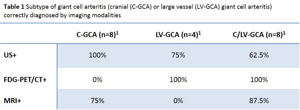
Results: In total, 42 patients with suspected GCA were included. Mean age was 70.7 years (SD 7.7) and 61.9% were female. The expert panel classified 23 patients as GCA after six months follow-up, of whom 95.7% met the revised ACR criteria for GCA. Specificity was 100% for US, FDG-PET/CT and MRI. Sensitivity was 69.6% for US, 52.2% for FDG-PET/CT and 56.5% for MRI (Figure 1). FDG-PET/CT results were positive for GCA in all patients with extracranial artery involvement (n=12), while US and MRI performed more optimal for isolated cranial artery involvement (n=8) (100% and 75% respectively) (Table 1).

Conclusion: The diagnostic value of US, FDG-PET/CT and MRI was comparable with high specificity and moderate sensitivity. FDG-PET/CT performed best in diagnosing extracranial artery involvement while US and MRI performed better for isolated cranial artery involvement. Diagnostic choices should be made considering availability of (state-of-the-art) equipment, operator experience, costs and suspected subtype. As US is non-invasive and allows quick visualization, this can be performed as a first test conform EULAR recommendations [1]. However, with high pre-test probability and inconclusive or negative results, further testing is required based on suspected subtypes.

[1] Dejaco C, Ramiro S, Duftner C, Besson FL, Bley TA, Blockmans D, Brouwer E, Cimmino MA, Clark E, Dasgupta B, Diamantopoulos AP, Direskeneli H, Iagnocco A, Klink T, Neill L, Ponte C, Salvarani C, Slart RHJA, Whitlock M, Schmidt WA. EULAR recommendations for the use of imaging in large vessel vasculitis in clinical practice. Ann Rheum Dis. 2018 May;77(5):636-643.

Supporting image 1

Figure 1: Diagnostic value in terms of sensitivity, specificity, positive predictive value and negative predictive value for three imaging modalities in patients with suspected giant cell arteritis. US=ultrasound, FDG-PET/CT=[18F]fluorodeoxyglucose-positron emission tomography/computed tomography, MRI=magnetic resonance imaging.

Supporting image 2

(1) Based on all imaging results in patients with a positive reference diagnosis. +=positive for giant cell arteritis. N=3 had negative results on all imaging modalities and could therefore not be classified in subtypes.


Disclosures: M. van Nieuwland, None; M. Vermeer, None; E. Colin, None; N. Wagenaar, None; O. Vijlbrief, None; J. van Zandwijk, None; R. Slart, Siemens Healthineers; H. Koffijberg, None; E. Groot Jebbink, None; E. Brouwer, None; D. Boumans, None; C. Alves, None.

To cite this abstract in AMA style:

van Nieuwland M, Vermeer M, Colin E, Wagenaar N, Vijlbrief O, van Zandwijk J, Slart R, Koffijberg H, Groot Jebbink E, Brouwer E, Boumans D, Alves C. Direct Comparison of Ultrasound, [18F]Fluorodeoxyglucose-Positron Emission Tomography/Computed Tomography and Magnetic Resonance Imaging: Early Diagnostics in Patients Suspected of Giant Cell Arteritis [abstract]. Arthritis Rheumatol. 2022; 74 (suppl 9). https://acrabstracts.org/abstract/direct-comparison-of-ultrasound-18ffluorodeoxyglucose-positron-emission-tomography-computed-tomography-and-magnetic-resonance-imaging-early-diagnostics-in-patients-suspected-of-giant-cell-arteriti/. Accessed .
  • Tweet
  • Click to email a link to a friend (Opens in new window) Email
  • Click to print (Opens in new window) Print

« Back to ACR Convergence 2022

ACR Meeting Abstracts - https://acrabstracts.org/abstract/direct-comparison-of-ultrasound-18ffluorodeoxyglucose-positron-emission-tomography-computed-tomography-and-magnetic-resonance-imaging-early-diagnostics-in-patients-suspected-of-giant-cell-arteriti/

Advanced Search

Your Favorites

You can save and print a list of your favorite abstracts during your browser session by clicking the “Favorite” button at the bottom of any abstract. View your favorites »

All abstracts accepted to ACR Convergence are under media embargo once the ACR has notified presenters of their abstract’s acceptance. They may be presented at other meetings or published as manuscripts after this time but should not be discussed in non-scholarly venues or outlets. The following embargo policies are strictly enforced by the ACR.

Accepted abstracts are made available to the public online in advance of the meeting and are published in a special online supplement of our scientific journal, Arthritis & Rheumatology. Information contained in those abstracts may not be released until the abstracts appear online. In an exception to the media embargo, academic institutions, private organizations, and companies with products whose value may be influenced by information contained in an abstract may issue a press release to coincide with the availability of an ACR abstract on the ACR website. However, the ACR continues to require that information that goes beyond that contained in the abstract (e.g., discussion of the abstract done as part of editorial news coverage) is under media embargo until 10:00 AM ET on November 14, 2024. Journalists with access to embargoed information cannot release articles or editorial news coverage before this time. Editorial news coverage is considered original articles/videos developed by employed journalists to report facts, commentary, and subject matter expert quotes in a narrative form using a variety of sources (e.g., research, announcements, press releases, events, etc.).

Violation of this policy may result in the abstract being withdrawn from the meeting and other measures deemed appropriate. Authors are responsible for notifying colleagues, institutions, communications firms, and all other stakeholders related to the development or promotion of the abstract about this policy. If you have questions about the ACR abstract embargo policy, please contact ACR abstracts staff at [email protected].

Wiley

  • Online Journal
  • Privacy Policy
  • Permissions Policies
  • Cookie Preferences

© Copyright 2025 American College of Rheumatology