Session Information
Session Type: Abstract Submissions (ACR)
Background/Purpose: Patients can fulfil the ASAS (Assessment of SpondyloArthritiS) sets of criteria with either objective evidence of sacroiliac joint damage (X-Ray) or inflammation (MRI) (“imaging” arm) or without such imaging abnormalities in B27 patients (“clinical” arm). Currently, there is still a debate regarding the imaging abnormalities permitting to classify a patient as fulfilling the imaging arm. The aim of the study was to evaluate different imaging abnormalities (e.g.inflammatory and chronic changes at the sacroiliac joint (SIJ) and spine level) as of potential interest for classification.
Methods: Prospective, multi-centre, observational study. 708 patients with early inflammatory back pain suggestive of spondyloarthritis were included in the DESIR cohort. Data collected: demographics, items of the ASAS criteria, disease activity, severity, quality of life. Inflammatory and chronic changes evaluated using either X-rays or MRI at both the SIJ and spine level by the radiologist/rheumatologist of the participating center. Statistical analysis: patients fulfilling the ASAS criteria were split in two groups (imaging vs. clinical) and within the clinical arm, with regard to the presence of CRP abnormality (e.g. >6mg/L). The other imaging abnormalities suggestive of SpA (e.g. MRI chronic changes of the SIJ, MRI inflammatory and chronic changes at the spine level, and the presence of at least 1 syndesmophyte at the cervical or lumbar level) present in each of the groups were depicted.
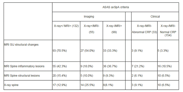
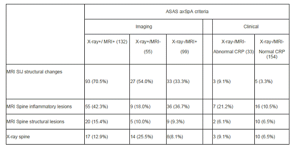
Results: 476 (69.8%) of the 708 included patients fulfilled the ASAS criteria, either the imaging (296 (60.1%)) or the clinical (190 (39.9%)) arms. Imaging findings different than the ones of the “imaging” definition of the ASAS criteria were observed (MRI-SIJ chronic changes (53.5% vs. 4.3%), MRI-Spine inflammatory changes (35.0% vs. 12.3%), MRI-spine chronic changes (11.9% vs. 6.4%) and X-ray-syndesmophytes (13.6% vs. 7.0%) in the imaging versus clinical arm, respectively.
The table summarizes the different imaging abnormalities observed.
MRI chronic changes of the SIJ were, as expected, more frequently observed in the subgroup of patients with X-ray damage of the SIJ. MRI inflammatory changes of the spine were more frequently observed in presence of other markers of inflammation. In the subgroups of patients without imaging abnormalities but abnormal CRP as much as 9.1% of patients presented with definite X-ray damage of the spine.
Conclusion: This study shows that different imaging abnormalities suggestive of SpA (e.g. MRI chronic changes of the SIJ) are present in early SpA, but need to be confirmed by central reading. Descriptive studies of these abnormalities in healthy controls are necessary to confirm (or not) whether these findings might be may be considered for classification in the imaging arm of the ASAS criteria.
Disclosure:
A. Moltó,
None;
S. Paternotte,
None;
D. van der Heijde,
None;
P. Claudepierre,
None;
M. Rudwaleit,
None;
M. Dougados,
None.
« Back to 2013 ACR/ARHP Annual Meeting
ACR Meeting Abstracts - https://acrabstracts.org/abstract/different-imaging-abnormalities-suggestive-of-spondyloarthritis-are-present-in-early-axial-spondyloarthritis-data-from-the-desir-cohort/


