Session Information
Session Type: ACR Poster Session A
Session Time: 9:00AM-11:00AM
Background/Purpose: Osteoarthritis (OA) of the first metatarsophalangeal joint (MTPJ) of the foot is the most common form of foot OA. The condition is typically evaluated using plain film radiographs, however magnetic resonance imaging (MRI) may provide more detailed insights into the disease process. Therefore, the purpose of this study was to develop a standardised atlas of MRI features of first MTPJ OA and assess its reliability.
Methods: We selected representative images covering the spectrum of OA severity from a database of first MTPJ OA for the following features: osteophytes (dorsal and plantar metatarsal head and dorsal proximal phalanx), joint space narrowing (first MTPJ and first metatarsal-sesamoid joint), bone marrow lesions (first metatarsal, proximal phalanx and sesamoids), cysts (first metatarsal and proximal phalanx), effusion (dorsal and plantar), capsular thickening (dorsal and plantar) and cartilage loss. Thirty cases were then independently scored with the atlas by two raters (SEM and HBM) to determine inter-rater reliability. Statistical analysis was conducted using percentage agreement and Gwet’s AC1 modification of the weighted kappa statistic (Ƙw) and were interpreted using the cut-offs proposed by Cohen (0-0.20 slight, 0.21-0.40 fair, 0.41-0.60 moderate, 0.61-0.80 substantial, 0.81-1.00 almost perfect). An a priori decision was made to exclude any observations with Ƙw <0.40 from the atlas. Reliability of a combined score summing all observations was also assessed using the intra-class correlation coefficient (ICC).
Results: An example MRI from a participant with first MTPJ OA is shown in the figure, and inter-rater reliability results are presented in the table. Percentage agreement ranged from 57 to 96%, and Ƙw scores ranged from 0.13 to 0.91. Of the 15 features documented with the atlas, 14 (93%) demonstrated acceptable reliability. Only plantar capsular thickening did not reach the reliability threshold, with a Ƙw of 0.13. The ICC for the combined score was 0.89 (95% CI 0.78 – 0.95), and after excluding plantar capsular thickening, the ICC was 0.90 (95% CI 0.80 – 0.95).
Conclusion: With the exception of first MTPJ plantar capsular thickening, MRI features of first MTPJ OA can be reliably documented using our standardised atlas. The use of this atlas will assist in documenting the severity of first MTPJ OA for epidemiological studies and for evaluating the effects of treatment in clinical trials.
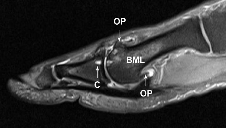
Figure. Example MRI of first MTPJ OA, demonstrating osteophytes (OP), cyst (C) and bone marrow lesion (BML).
Table. Reliability results.
|
Variable |
% agreement |
Gwet’s AC1 Ƙw |
Interpretation |
|
Osteophytes, dorsal metatarsal head1 |
91 |
0.72 (0.53 – 0.91) |
Substantial |
|
Osteophytes, plantar metatarsal head1 |
94 |
0.81 (0.68 – 0.94) |
Almost perfect |
|
Osteophytes, dorsal proximal phalanx1 |
86 |
0.54 (0.24 – 0.84) |
Moderate |
|
Joint space narrowing, 1st MTPJ1 |
95 |
0.88 (0.82 – 0.94) |
Almost perfect |
|
Joint space narrowing, 1st metatarsal-sesamoid2 |
80 |
0.60 (0.30 – 0.91) |
Moderate |
|
Bone marrow lesions, metatarsal3 |
96 |
0.87 (0.80 – 0.94) |
Almost perfect |
|
Bone marrow lesions, proximal phalanx3 |
96 |
0.87 (0.76 – 0.98) |
Almost perfect |
|
Bone marrow lesions, sesamoids2 |
67 |
0.40 (0.03 – 0.77) |
Fair |
|
Cysts, metatarsal2 |
90 |
0.81 (0.58 – 1.00) |
Almost perfect |
|
Cysts, proximal phalanx2 |
93 |
0.91 (0.78 – 1.00) |
Almost perfect |
|
Effusion – dorsal2 |
83 |
0.76 (0.53 – 0.99) |
Substantial |
|
Effusion – plantar2 |
80 |
0.67 (0.39 – 0.95) |
Substantial |
|
Thickening – dorsal2 |
70 |
0.40 (0.05 – 0.75) |
Fair |
|
Thickening – plantar2 |
57 |
0.13 (-0.24 – 0.51) |
Slight |
|
Cartilage loss2 |
80 |
0.60 (0.29 – 0.91) |
Moderate |
|
1 scored as none, mild, moderate or severe 2 scored as present or absent 3 scored as none, <25%, 25-50%, >50% |
|||
To cite this abstract in AMA style:
Munteanu S, Landorf K, Tan J, Auhl M, Allan J, Buldt A, Menz HB. Development of a Magnetic Resonance Imaging Atlas for the Classification of Osteoarthritis of the First Metatarsophalangeal Joint [abstract]. Arthritis Rheumatol. 2018; 70 (suppl 9). https://acrabstracts.org/abstract/development-of-a-magnetic-resonance-imaging-atlas-for-the-classification-of-osteoarthritis-of-the-first-metatarsophalangeal-joint/. Accessed .« Back to 2018 ACR/ARHP Annual Meeting
ACR Meeting Abstracts - https://acrabstracts.org/abstract/development-of-a-magnetic-resonance-imaging-atlas-for-the-classification-of-osteoarthritis-of-the-first-metatarsophalangeal-joint/