Session Information
Date: Monday, November 9, 2020
Session Type: Abstract Session
Session Time: 5:00PM-5:50PM
Background/Purpose: Conventional radiography of the sacroiliac joints is still recommended as the first imaging method if axial spondyloarthritis (axSpA) is suspected. Furthermore, radiographic sacroiliitis is included – together with sacroiliitis on magnetic resonance imaging – in the Assessment of Spondyloarthritis International Society (ASAS) classification criteria for axSpA. Depending on the presence or absence of definite radiographic sacroiliitis, axSpA can be classified either as radiographic axSpA (r-axSpA, synonymous to ankylosing spondylitis) or non-radiographic axSpA (nr-axSpA). Although conventional radiography of sacroiliac joints still plays an important role in clinical practice and for clinical trials, the reliability of radiographic sacroiliitis assessment has been reported in a number of studies as mostly poor, even if performed by expert readers. Furthermore, it was shown that untrained local readers perform worse than expert readers specialised in SpA. One possible solution to achieve a comparable high accuracy as an expert on detection of radiographic sacroiliitis, even in non-specialised clinics, could be development of an artificial intelligence-based model for analysis of radiographs.
The aim of this study was to develop and validate an artificial neural network for the detection of definite radiographic sacroiliitis as a manifestation of axial spondyloarthritis.
Methods: Conventional radiographs of sacroiliac joints from two independent cohorts of patients with axSpA were used. The first cohort (PROOF) consisted of 1669 radiographs and was used for training and validation of a neural network. The second cohort consisted of 100 randomly selected radiographs from GESPIC, which were used as an independent test dataset. In both cohorts all radiographs underwent central reading; the final decision on the presence or absence of definite radiographic sacroiliitis according to the radiographic criterion of the modified New York criteria was used as a reference. For performance evaluation of the neural network, areas under the receiver operating characteristic curves (AUROC) were calculated. Sensitivity and specificity for the prediction cut-offs were calculated. Cohen’s Kappa and the absolute agreement were used to assess the agreement between the neural network and the human readers.
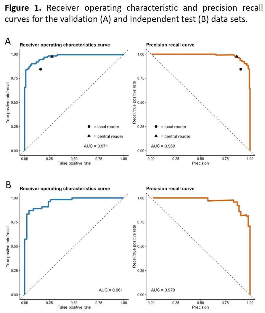
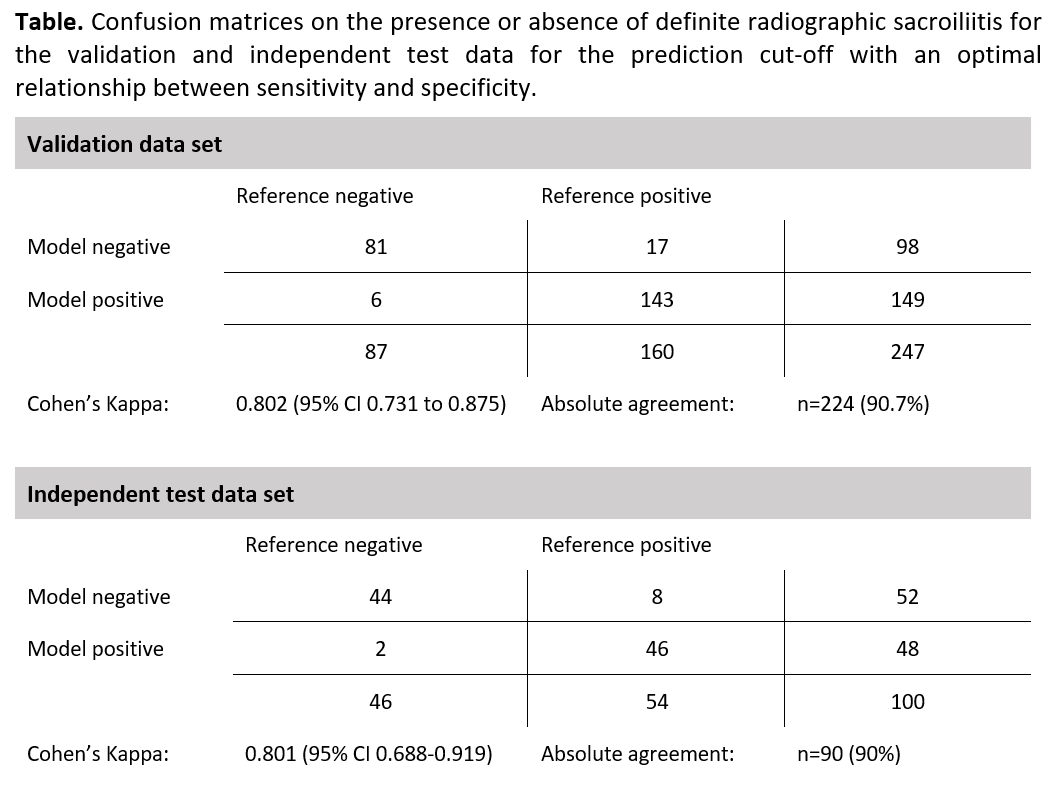
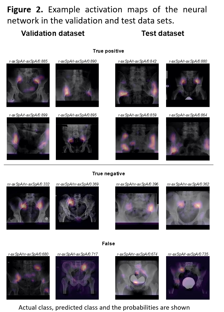
Results: The neural network achieved an excellent performance in recognition of definite radiographic sacroiliitis with AUROC of 0.97 and 0.96 for the validation and test datasets, respectively (Figure 1). Sensitivity and specificity for the cut-off weighting both measurements equally were 0.90 and 0.93 for the validation and 0.87 and 0.97 for the test set. The Cohen’s kappa between the neural network and the reference judgements were 0.80 for both validation and test sets, and the absolute agreement on the classification yielded 91% and 90%, respectively (Table). Examples of the activation maps of the neural network are presented in Figure 2.
Conclusion: Artificial neural networks enable the accurate detection of definite radiographic sacroiliitis relevant for the diagnosis and classification of axSpA.
To cite this abstract in AMA style:
Bressem K, Vahldiek J, Adams L, Niehues S, Haibel H, Rios Rodriguez V, Torgutalp M, Protopopov M, Proft F, Rademacher J, Sieper J, Rudwaleit M, Hamm B, Makowski M, Hermann K, Poddubnyy D. Development and Validation of an Artificial Intelligence Approach for the Detection of Radiographic Sacroiliitis [abstract]. Arthritis Rheumatol. 2020; 72 (suppl 10). https://acrabstracts.org/abstract/development-and-validation-of-an-artificial-intelligence-approach-for-the-detection-of-radiographic-sacroiliitis/. Accessed .« Back to ACR Convergence 2020
ACR Meeting Abstracts - https://acrabstracts.org/abstract/development-and-validation-of-an-artificial-intelligence-approach-for-the-detection-of-radiographic-sacroiliitis/


