Session Information
Session Type: Abstract Submissions (ACR)
Background/Purpose: Cartilage morphometry on magnetic resonance images (MRIs) may become an acceptable outcome measure for clinical trials among patients with knee osteoarthritis (OA). However, obtaining accurate and reproducible cartilage data is burdensome. To conduct large clinical trials it will be vital to use an efficient, sensitive and reproducible method to assess cartilage morphometry. The purpose of this study was to develop and validate a rapid semi-automated method to detect cartilage loss in the medial tibiofemoral compartment.
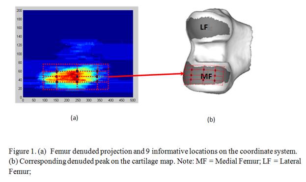
Methods: The rapid knee cartilage quantification method was developed using 263 manually-segmented knee MRIs. We designed a pair of two dimensional, rectangular, universal coordinate systems to represent the articular surface of the distal medial femur and proximal medial tibia. Next, we projected segmented cartilage onto coordinate system and evenly selected 9 informative locations across the region most frequently denuded (Figure 1). The primary outcome is a cartilage damage index (CDI), which summates the products of cartilage thickness, cartilage length and voxel size from each informative location. To validate CDI we selected 102 subjects from Osteoarthritis Initiative with a diverse range of Kellgren-Lawrence (KL) grades (0 to 4). 3D Double-echo steady-state sagittal images were obtained on four 3-Tesla systems (0.37 mm x 0.37 mm, 0.7 mm slice thickness). One reader used customized software to measure the CDI in the medial femur and tibia from the baseline and 24-month visit. Another reader evaluated 20 subjects to establish the inter-tester reliability test.
Results: The average measurement time was 14 minutes per pair of knees. The intra-tester reliability (intraclass correlation coefficient ICC [3,1 model] 0.95 to 0.99) and inter-tester reliability (ICC [2,1 model] 0.85 to 0.94) were good. Knees with greater medial joint space narrowing (JSN) had lower mean CDI (i.e. greater loss, Table 1). Knees with radiographic progression (JSN grade change) had 4.5 to 22 times greater CDI loss than knees with no progression (p < 0.0001), Table 2.
Conclusion: This novel knee cartilage damage quantification method is rapid, reliable, and has good construct validity in the medial tibiofemoral compartment. It has utility for deployment in large epidemiological studies.
|
||||||||||||||||||||||||||||||||||||||||||||||||||||||||||||||||||||||||
|
Table 2.Change in cartilage damage index among knees with and without structural progression defined by change in radiographic JSN grade |
||||||||||||||||||||||||||||||||||||||||||||||||||||||||||||||||||||||||
|
Cartilage Measure |
JSN change |
N |
Mean |
SD |
SRM |
p-value |
||||||||||||||||||||||||||||||||||||||||||||||||||||||||||||||||||
|
Femur CDI (Change)
|
No change |
74 |
-3.38 |
47.98 |
-0.07 |
<0.0001 |
||||||||||||||||||||||||||||||||||||||||||||||||||||||||||||||||||
|
Change |
25 |
-66.17 |
56.76 |
-1.17 |
||||||||||||||||||||||||||||||||||||||||||||||||||||||||||||||||||||
|
Tibia CDI (Change)
|
No change |
74 |
-12.85
|
23.38 |
-0.55 |
<0.0001 |
||||||||||||||||||||||||||||||||||||||||||||||||||||||||||||||||||
|
Change |
25 |
-55.55 |
39.82 |
-1.40 |
||||||||||||||||||||||||||||||||||||||||||||||||||||||||||||||||||||
|
Tibiofemoral CDI (Change)
|
No change |
74 |
-16.23 |
56.68 |
-0.29 |
<0.0001 |
||||||||||||||||||||||||||||||||||||||||||||||||||||||||||||||||||
|
Change |
25 |
-121.70 |
77.17 |
-1.58 |
||||||||||||||||||||||||||||||||||||||||||||||||||||||||||||||||||||
|
Notes: JSN change, the follow-up has different JSN score with baseline; N = number of knees (the numbers were excluded JSN=3 which is the highest grade); SD = standard deviation; SRM = standard response mean |
||||||||||||||||||||||||||||||||||||||||||||||||||||||||||||||||||||||||
Disclosure:
M. Zhang,
None;
J. B. Driban,
None;
L. L. Price,
None;
D. Harper,
None;
G. H. Lo,
None;
E. Miller,
None;
R. J. Ward,
None;
T. E. McAlindon,
None.
« Back to 2013 ACR/ARHP Annual Meeting
ACR Meeting Abstracts - https://acrabstracts.org/abstract/development-and-validation-of-a-rapid-knee-cartilage-quantification-method/