Session Information
Date: Wednesday, October 24, 2018
Title: 6W020 ACR Abstract: Metabolic & Crystal Arthropathies–Basic Science & Imaging (2964–2969)
Session Type: ACR Concurrent Abstract Session
Session Time: 11:00AM-12:30PM
Background/Purpose:
Many recent studies have shown an association between gout and increased cardiovascular risk, however the mechanism by which this occurs is unclear. Dual-Energy Computed Tomography (DECT) is a new technology that has been used to detect subclinical monosodium urate (MSU) deposition in the joints of patients with gout. In this study, we investigated whether DECT could also detect MSU deposition in the vasculature of patients with gout.
Methods:
Fourteen tophaceous gout patients and ten non-tophaceous patients were recruited from a single-center urban academic hospital. All patients underwent DECT scans of the neck and chest using a Siemens Somatom Force CT scanner, and images were processed with SyngoVia imaging software to identify MSU deposits. An automated volume assessment program was used to calculate the volume of urate deposits in the vasculature of patients.
Results:
The range of uric acid deposition within patients’ vasculature varied widely, from 0 mm3 to over 450 mm3, but the majority of study participants (88%) were found to have evidence of MSU deposition in their vasculature. Due to the large volume of these deposits, we did not feel that this was consistent with artifact. The average MSU deposition volumes were 109.4mm3 for the tophaceous patients and 116.5 mm3 for the non-tophaceous patients, but this difference was not statistically significant (p = 0.84).
Conclusion:
DECT is able to detect the presence of MSU deposition in the vasculature of patients with both tophaceous and non-tophaceous gout. Whether or not this results in cardiovascular inflammation remain to be determined. Future directions include using PET/MRI to evaluate for local inflammation at the site of MSU deposits in the vasculature.
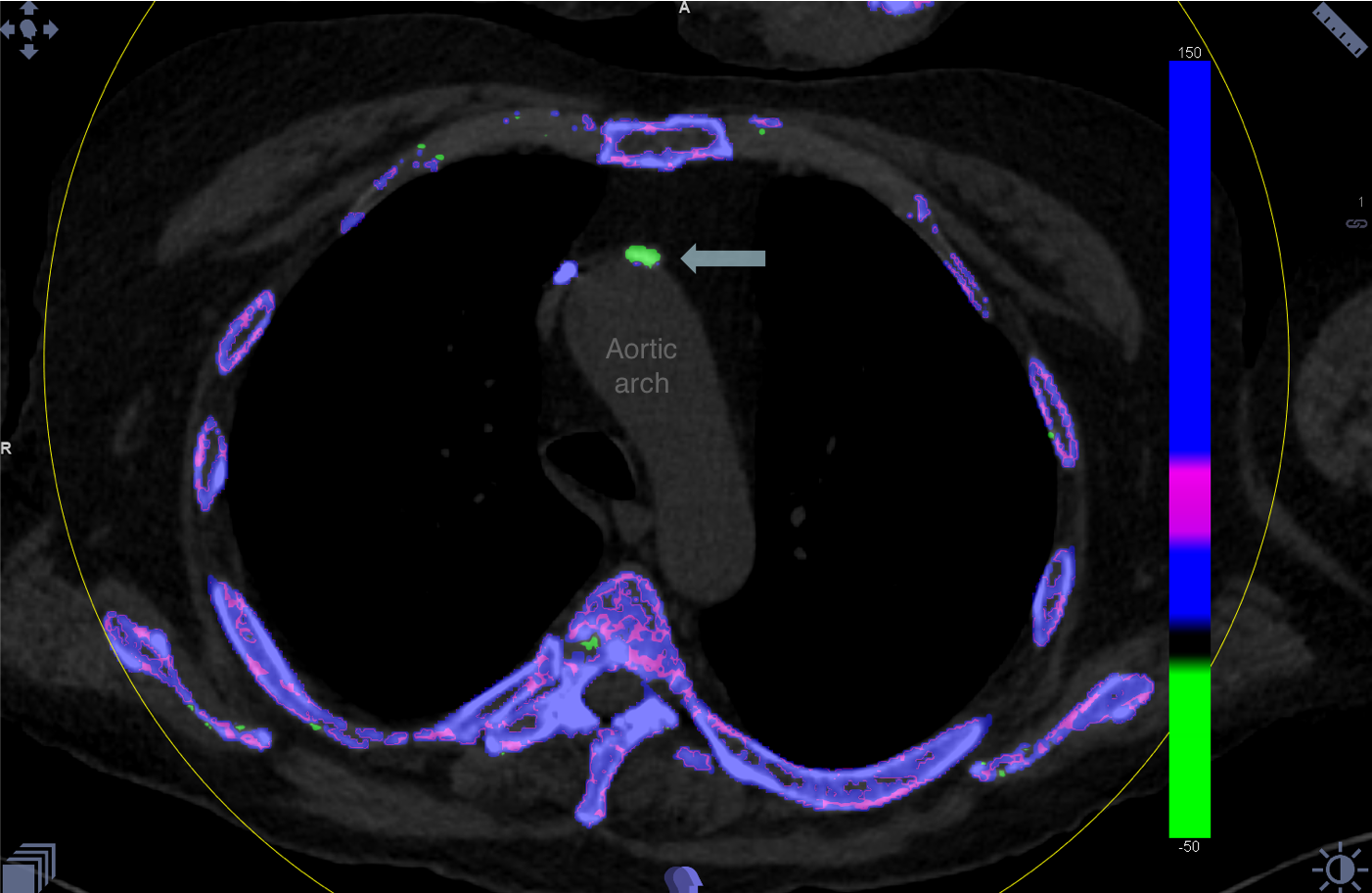
Figure 1: This patient’s aortic arch has about 20 mm2 of uric acid in this slide alone, seen in green.

Figure 2: This patient has multiple discrete spots of MSU crystallization (green), lining his ascending thoracic aorta and in the interior of his coronary artery.
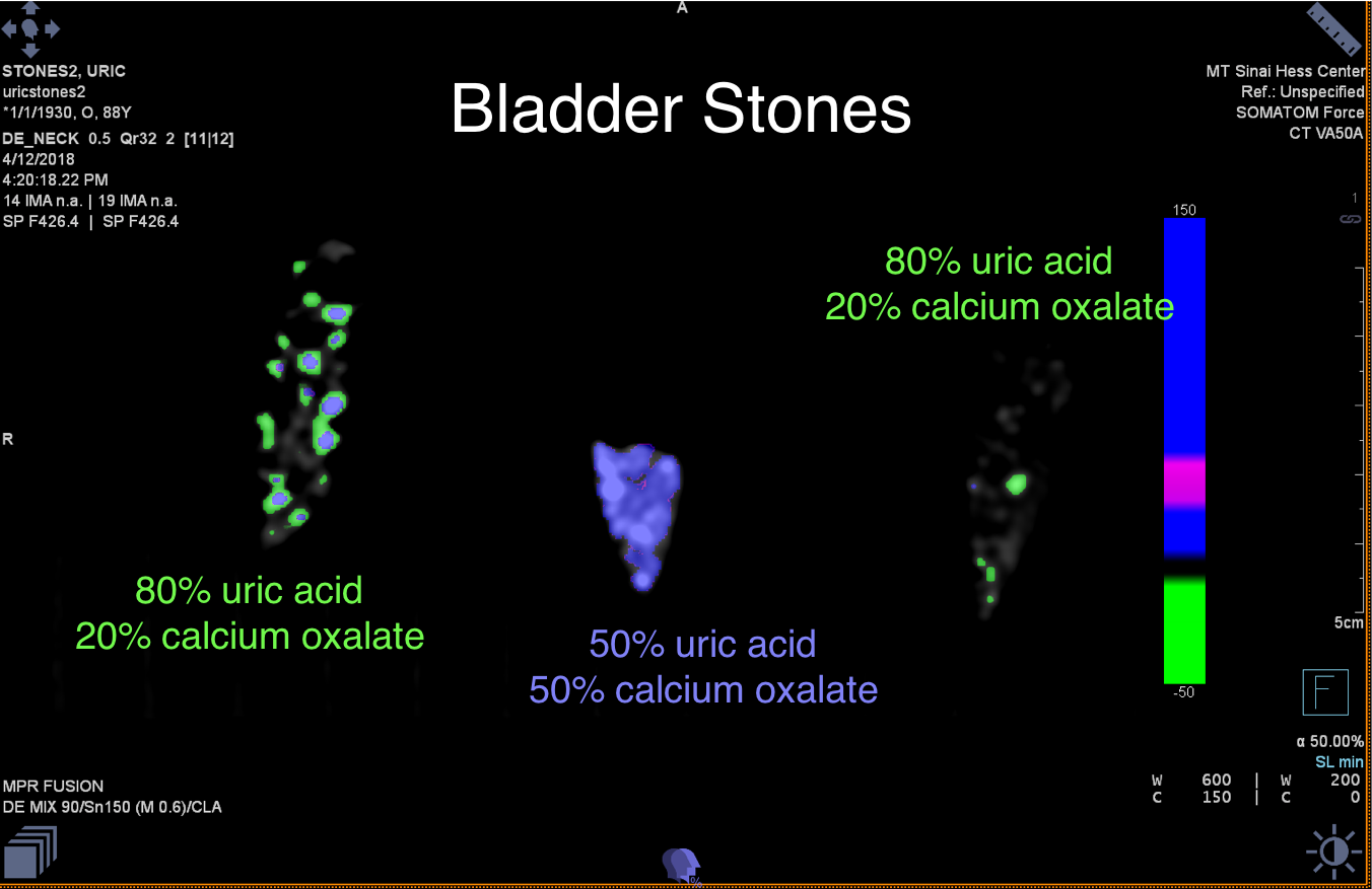
Figure 3: To confirm the calibration of the DECT scanner, we tested three bladder stone samples whose UA composition ranged from 50-80%; the remainder being calcium oxalate. Scans and analysis confirmed that the scanner is able to pick up MSU (green) and differentiate it from calcium derivatives (purple).
To cite this abstract in AMA style:
Barazani S, Chi W, Pyzik R, Jacobi A, O'Donnell T, Fayad Z, Mani V, Ali Y. Detection of Uric Acid Crystals in the Vasculature of Patients with Gout Using Dual-Energy Computed Tomography [abstract]. Arthritis Rheumatol. 2018; 70 (suppl 9). https://acrabstracts.org/abstract/detection-of-uric-acid-crystals-in-the-vasculature-of-patients-with-gout-using-dual-energy-computed-tomography/. Accessed .« Back to 2018 ACR/ARHP Annual Meeting
ACR Meeting Abstracts - https://acrabstracts.org/abstract/detection-of-uric-acid-crystals-in-the-vasculature-of-patients-with-gout-using-dual-energy-computed-tomography/