Session Information
Session Type: Abstract Submissions (ACR)
Background/Purpose
MicroRNAs (miRs) are an important class of regulators of gene expression that are associated with a variety of biological functions. Deregulation of endogenous miR155 was observed in many autoimmune conditions, including SLE. We herein examine the role of miR155 in the development of systemic manifestations in murine pristane induced lupus by evaluating the severity of organ involvement and assessing serum antibody-levels and T helper cell homeostasis.
Methods
MiR155-deficient (miR155-PIL) and C57/Bl6 (WT-PIL) mice were injected i.p. with 0.5ml of pristane or PBS as control (WT-PBS). In order to observe the effects of miR155 deficiency in fully developed SLE, we analyzed the mice 8 months after induction.
A blinded specialist appraised histological features of GN using the composite kidney biopsy score (KBS). Histological features of pneumonitis were quantified by an image analysis system: Lungs were scored for the severity of perivascular inflammation by analyzing the numbers of affected vessels and the area of the inflammatory infiltrate. In order to assess the composition of these infiltrates, specimens were stained with B220 (B), CD3 (T), Neu7/4 (neutrophils) and F4/80 (macrophages) and analyzed by cell-identification algorithms for nuclear segmentation (HistoQuest¨).
Lymphocytes were isolated from spleens and analyzed separately for each mouse by standard FACS procedures. For analysis of the Th1, 2 or 17 subsets, respectively, cells were re-stimulated in vivo with anti-CD3 and anti-CD28abs.
Results
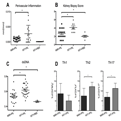
Lungs were affected in both pristane-treated groups, but not in controls. MiR155-PIL had reduced lupus severity as indicated by significantly decreased perivascular inflammatory area with B cells being the most prominent inflammatory cell type in the HistoQuest analysis (Fig. 1A). Without showing clinical abnormalities WT-PIL had a more severe renal involvement in the kidney biopsy score than miR-PIL (Fig. 1B). Corresponding with reduced severity in organ involvement, miR155-PIL had lower serum levels of anti-dsDNA-abs (Fig. 1C), decreased frequencies of CD4+ cells (14.24 ± 0.7587 vs. 18.04 ± 1.075, p=0.01) and slightly lower frequencies of activated CD4+CD25+Foxp3– cells (1.539 ± 0.1279 vs. 1.838 ± 0.2259, p=ns.). Interestingly, also frequencies of CD4+CD25+Foxp3+ regulatory T cells were lower in Mir155-PIL (1.689 ± 0.1388 vs. 2.375 ± 0.2320, p=0.03). Upon restimulation, CD4+ cells showed a more pronounced Th2 and Th17 response in WT-PIL, but no significant differences in Th1 phenotype (Fig. 1D).
Conclusion
MiR155 deficiency in PIL mice did not prevent the development of disease, but was associated with less severe lung and kidney involvement, lower serum auto-abs levels and lower Th17 and Th2 frequencies when analyzed in fully established PIL after 8 months. Thus, antagonisation of miR155 might be a beneficial future approach in treating SLE.
Disclosure:
H. Leiss,
None;
W. Salzberger,
None;
I. Gessl,
None;
B. Schwarzecker,
None;
N. Kozakowski,
None;
S. Blüml,
None;
A. Puchner,
None;
B. Niederreiter,
None;
C. W. Steiner,
None;
J. Smolen,
None;
G. H. Stummvoll,
None.
« Back to 2014 ACR/ARHP Annual Meeting
ACR Meeting Abstracts - https://acrabstracts.org/abstract/decreased-severity-of-pristane-induced-lupus-in-mir155-deficient-mice/

