Session Information
Date: Monday, November 9, 2015
Title: Spondylarthropathies and Psoriatic Arthritis - Comorbidities and Treatment Poster II
Session Type: ACR Poster Session B
Session Time: 9:00AM-11:00AM
Background/Purpose:
Quantifying syndesmophyte (Synd) length and angle in axial
spondyloarthritis (axSpA) patients (pts) can be a helpful tool in monitoring
disease progression. Objective was to evaluate in-house-developed software that
aids in quantifying Synds length and angle on radiographs, to test its inter and intrareader reliability and to assess
progression over time.
Methods:
The cervical and lumbar spine radiographs at baseline and
follow up (24 months) of 18 axSpA pts were analysed twice using the software
tool, by two independent readers, blinded for time sequence and the results of
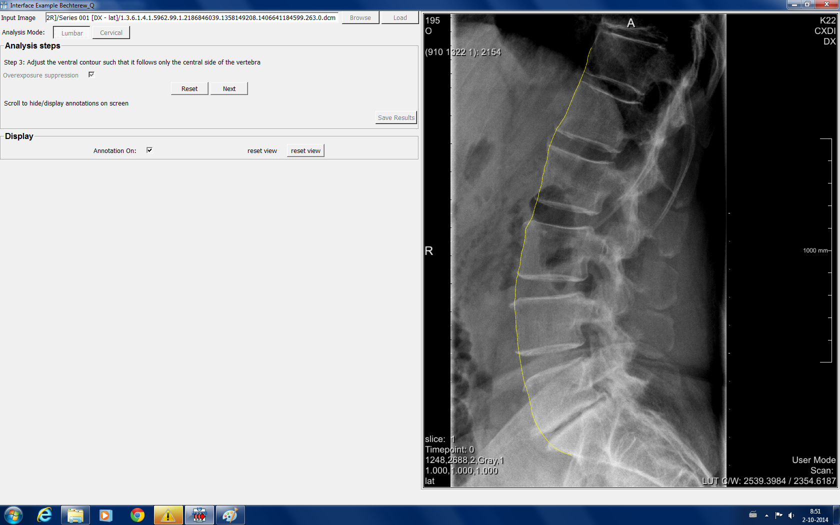
the other reader. The software establishes the vertebral body (VB) contours
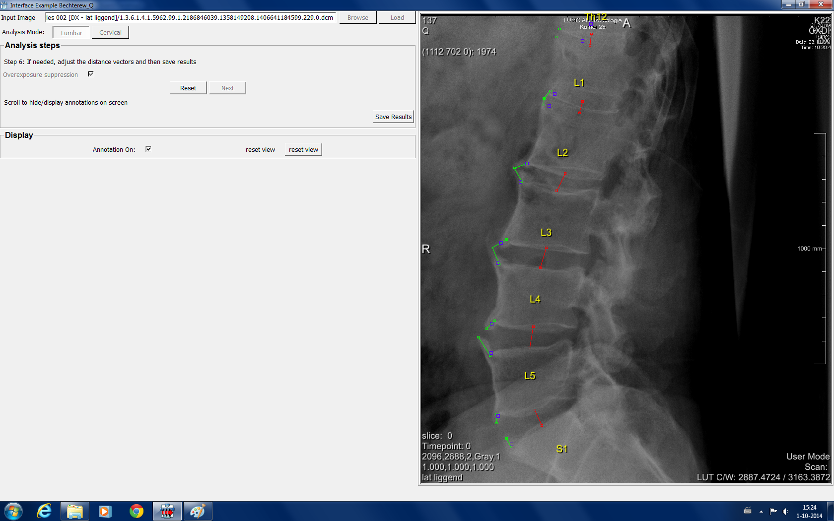
based on signal contrast and anatomical landmarks (figure 1). The length and
angle of Synds is based on the distance between the maximum extending point and
the ‘native’ VB corner (figure 2). Length was measured in mm and sum scores
were calculated per pt by adding up all Synds. Pts were also analysed using the
mSASSS. Inter- and intrareader reliability were
analysed using Bland Altman (B&A) plots. Reliability of progression was
evaluated using smallest detectable change (SDC) analysis.
Results:
In total, each reader analysed 1728 vertebral corners (VC) of
72 images. B&A plots for intra and interreader variability showed a large
variability in length of Synds in both the individual VCs and for the total sum
of the patients (figure 3). When analysing change score over time within one
patient, SDC showed that almost all progression fell within the SDC (SDC= 12.9
and 20.4 mm) (for reading 1: 11/18 patients (61%) and reading 2: 15/18 (83%)),
while those patients did show ‘real progression’ as measured with the mSASSS.
Conclusion:
Computer aided measurement of Synds on radiographs was not
feasible. The restrictions associated with radiographs (overprojection, patient
position dependent variability) and the size of the lesions
of interest were not compatible. Overall, semi-automatic detection did
not have added value to existing manual tools.
Figure 1
Figure 2
Figure 3
To cite this abstract in AMA style:
de Bruin F, van den Berg R, Reijnierse M, van der Heijde D, Stoel BC. Computer Aided Syndesmophyte Measurement on Spinal Radiographs in Axial Spondyloarthritis Patients [abstract]. Arthritis Rheumatol. 2015; 67 (suppl 10). https://acrabstracts.org/abstract/computer-aided-syndesmophyte-measurement-on-spinal-radiographs-in-axial-spondyloarthritis-patients/. Accessed .« Back to 2015 ACR/ARHP Annual Meeting
ACR Meeting Abstracts - https://acrabstracts.org/abstract/computer-aided-syndesmophyte-measurement-on-spinal-radiographs-in-axial-spondyloarthritis-patients/


