Session Information
Session Type: ACR Concurrent Abstract Session
Session Time: 9:00AM-10:30AM
Background/Purpose: A compensated polarizing microscopy has been used for detecting monosodium urate (MSU) or calcium pyrophosphate dihydrate (CPPD) crystals to confirm the diagnosis of gout or pseudogout for more than 50 years. However, accuracy of the methodology is affected by crystal concentration, time invested reading the slide and operator skill. As the procedure is only billable through a Clinical Laboratory Improvement Amendments (CLIA)-certified lab, its bedside use has waned over the years. There is a need for a more efficient and accurate method. Herein, we present a novel method to detect birefringent crystals in synovial fluid by designing a lens-free on-chip polarizing microscope.
Methods: Lens-free on-chip microscopy based on digital in-line holography is a novel imaging technique that has achieved wide field of view (>20 mm2) and high-resolution biomedical imaging in a simple, cost-effective and field-portable design. We modified this technology with a circular polarizer and an angle-mismatched analyzer so that the partially-coherent light that is passed through the birefringent crystals can be enhanced and detected by a digital image sensor. Two images of every sample are taken with the sample rotated by 90 degrees to eliminate any non-birefringent objects. An image processing algorithm and post image color-coding was applied to enhance the quality and to reproduce the similar color images to those of a compensated polarizing microscope. Imaging of CPPD crystals are in progress at the time of this abstract submission.
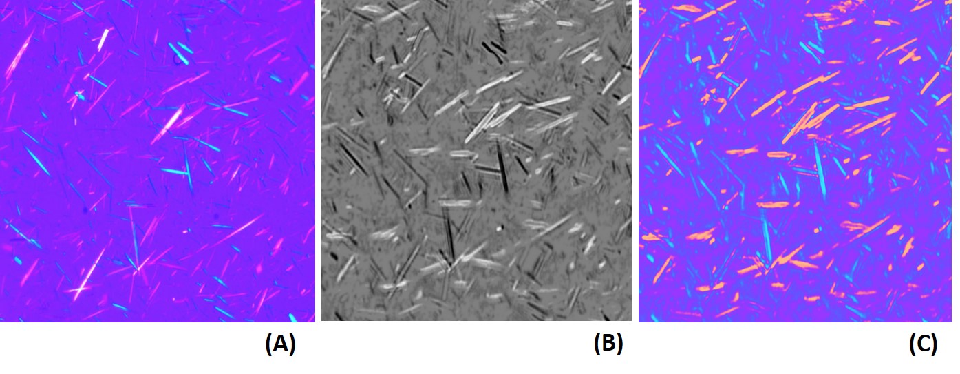
Results: Figure 1 shows the comparison of MSU crystal images taken by a compensated polarizing microscope (A), a lens-free differential grayscale image (B) and a lens-free color-coded image (C). The images taken by the novel technique was able to accurately demonstrate the direction and the strength of birefringence and the shape of MSU crystals. Two independent experts reviewed multiple images and determined that they were comparable to those from a conventional polarizing microscope.
Conclusion: We developed a digital lens-free on-chip polarizing microscope that can detect birefringent crystals over a wide field of view. It uses a computerized image analysis algorithm and a digital image sensor, which overcomes the weakness of time-consuming, operator-dependent property of the conventional polarizing microscope. Its time-efficiency, small size and low cost could make it more easily used in a daily practice, to facilitate synovial fluid examination for the diagnosis of crystal arthropathy. The process is currently completed for MSU crystals. Validation of CPPD crystal images taken by this novel technology is currently in progress.
Fig. 1: Conventional and Lens-Free On-Chip Polarizing Microscopy images of MSU crystals.
To cite this abstract in AMA style:
Lee SY, Zhang Y, Furst DE, Rosenthal A, Schumacher R, FitzGerald J, Ozcan A. Computational Polarizing Microscopy: A Novel Method to Detect Birefringent Crystals Using Lens-Free on-Chip Microscopy [abstract]. Arthritis Rheumatol. 2016; 68 (suppl 10). https://acrabstracts.org/abstract/computational-polarizing-microscopy-a-novel-method-to-detect-birefringent-crystals-using-lens-free-on-chip-microscopy/. Accessed .« Back to 2016 ACR/ARHP Annual Meeting
ACR Meeting Abstracts - https://acrabstracts.org/abstract/computational-polarizing-microscopy-a-novel-method-to-detect-birefringent-crystals-using-lens-free-on-chip-microscopy/

