ACR Meeting Abstracts

ACR Meeting Abstracts

  • Meetings
    • ACR Convergence 2024
    • ACR Convergence 2023
    • 2023 ACR/ARP PRSYM
    • ACR Convergence 2022
    • ACR Convergence 2021
    • ACR Convergence 2020
    • 2020 ACR/ARP PRSYM
    • 2019 ACR/ARP Annual Meeting
    • 2018-2009 Meetings
    • Download Abstracts
  • Keyword Index
  • Advanced Search
  • Your Favorites
    • Favorites
    • Login
    • View and print all favorites
    • Clear all your favorites
  • ACR Meetings

Abstract Number: 1043

Comparison of Optical and Ultrasound Imaging in Lupus Arthritis

Wei Tang1, Alessandro Marone2, Leila Khalili1, Xin Wang1, Shane Murray1, Sean Inzerillo1, Moegammad Bardien2, Vedant Gaikwad2, Stephen Kim2, Laura Geraldino-Pardilla1, Andreas Hielscher2 and Anca Askanase3, 1Division of Rheumatology, Columbia University Irving Medical Center, New York, NY, 2Department of Biomedical Engineering, Tandon School of Engineering, New York University, Brooklyn, NY, 3Columbia University Medical Center, New York, NY

Meeting: ACR Convergence 2023

Keywords: Imaging, Systemic lupus erythematosus (SLE)

  • Tweet
  • Click to email a link to a friend (Opens in new window) Email
  • Click to print (Opens in new window) Print
Session Information

Date: Monday, November 13, 2023

Title: (1033–1051) Imaging of Rheumatic Diseases Poster I

Session Type: Poster Session B

Session Time: 9:00AM-11:00AM

Background/Purpose: Almost all patients with SLE experience joint problems and arthritis, 12% suffer permanent joint damage. The wide variability in SLE arthritis and the limitations of existing assessment tools make it difficult to identify musculoskeletal involvement in clinical care and clinical trials. Identifying lupus arthritis based on physical exam remains challenging. Clinicians need a robust, easy-to-use imaging technique to diagnose and monitor arthritis in SLE. Ultrasound (US) and magnetic resonance (MRI) allow for more objective assessments but are plagued by operator dependence and high cost. With the support of a DoD award, we are developing a prototype imaging system that uses lasers and light detectors to evaluate finger joints in SLE. Here we describe preliminary data on comparisons between optical imaging and US evaluations of affected and unaffected proximal interphalangeal (PIP) joints.

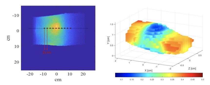
Methods: Fifteen SLE patients were evaluated, 8 PIP joints (bilateral PIP 2-5)/patient were imaged, with a total of 120 images. A systematic multiplanar grey-scale (GS) and power doppler (PD) examination of PIP joints was performed using the Outcome Measures in Rheumatology (OMERACT) consensus definitions for joint pathologies, and a combined PDGS score was calculated. Immediately after the US, joints were examined using optical imaging. The instrumentation uses a 670nm, 8mW laser with a 1 mm diameter beam modulated at 300 MHz as light source. The laser beam is focused on the dorsal surface of each finger and scans across the PIP joints (as shown in Figure 1, left). The transmitted light is collected by an intensified charge-coupled device camera and the absorption and scattering distributions inside the finger are reconstructed (Fig.1 shows absorption) using a radiative transfer equation model.

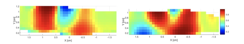
Results: The PDGS scored 34 joints 0, 44 – 1, 20 – 2 and 22 – 3. The absorption and scattering data were obtained from the reconstructed images. Figure 2 shows a cross section through an unaffected joint (PDGS=0) on the left and an affected joint (PDGS=3) on the right. The average absorption coefficient of joints with PDGS = 0 or 1 was 0.52±0.19 cm-1, while joints with PDGS = 2 or 3 the absorption was 0.60±0.19 cm-1, p < 0.05. Similarly, we found that the difference in the scattering coefficients between the two groups was also statistically significant (10.8±1.9 cm-1 versus 11.8±2.1 cm-1, p< 0.01).

Conclusion: Optical imaging correlated with PDGS findings in 120 evaluated lupus arthritis PIP joints. Further examination of SLE arthritis using the new US OMERACT criteria that includes a tenosynovitis score and MRI will allow for better definitions of PIP arthritis and improve the correlations with optical imaging. The system, which could take the form of an ergonomic glove, will allow the investigators to image lupus arthritis, both in the office and at home, to diagnose and monitor arthritis progress. The goal is to create a portable, wearable, imaging device that permits real-time monitoring of SLE arthritis to improve patient care and outcomes.

Supporting image 1

Figure 1. Left, example of the collected raw signal from the system, with the red dots indicating the different positions of the laser during the acquisition protocol. Right, example of a three-dimensional reconstruction of a joint’s absorption properties.

Supporting image 2

Figure 2. Representative cross-sectional images of “unaffected” (PDGS=0, left) and “affected” (PDGS=3, right) PIP joints absorption properties.


Disclosures: W. Tang: None; A. Marone: None; L. Khalili: None; X. Wang: None; S. Murray: None; S. Inzerillo: None; M. Bardien: None; V. Gaikwad: None; S. Kim: None; L. Geraldino-Pardilla: None; A. Hielscher: None; A. Askanase: AbbVie, 2, Amgen, 2, AstraZeneca, 2, Aurinia, 2, Bristol-Myers Squibb, 2, Celgene, 2, Eli Lilly, 2, Genentech, 2, GSK, 2, Idorsia, 2, Janssen, 2, Mallinckrodt, 2, Pfizer, 2, UCB Pharma, 2.

To cite this abstract in AMA style:

Tang W, Marone A, Khalili L, Wang X, Murray S, Inzerillo S, Bardien M, Gaikwad V, Kim S, Geraldino-Pardilla L, Hielscher A, Askanase A. Comparison of Optical and Ultrasound Imaging in Lupus Arthritis [abstract]. Arthritis Rheumatol. 2023; 75 (suppl 9). https://acrabstracts.org/abstract/comparison-of-optical-and-ultrasound-imaging-in-lupus-arthritis/. Accessed .
  • Tweet
  • Click to email a link to a friend (Opens in new window) Email
  • Click to print (Opens in new window) Print

« Back to ACR Convergence 2023

ACR Meeting Abstracts - https://acrabstracts.org/abstract/comparison-of-optical-and-ultrasound-imaging-in-lupus-arthritis/

Advanced Search

Your Favorites

You can save and print a list of your favorite abstracts during your browser session by clicking the “Favorite” button at the bottom of any abstract. View your favorites »

All abstracts accepted to ACR Convergence are under media embargo once the ACR has notified presenters of their abstract’s acceptance. They may be presented at other meetings or published as manuscripts after this time but should not be discussed in non-scholarly venues or outlets. The following embargo policies are strictly enforced by the ACR.

Accepted abstracts are made available to the public online in advance of the meeting and are published in a special online supplement of our scientific journal, Arthritis & Rheumatology. Information contained in those abstracts may not be released until the abstracts appear online. In an exception to the media embargo, academic institutions, private organizations, and companies with products whose value may be influenced by information contained in an abstract may issue a press release to coincide with the availability of an ACR abstract on the ACR website. However, the ACR continues to require that information that goes beyond that contained in the abstract (e.g., discussion of the abstract done as part of editorial news coverage) is under media embargo until 10:00 AM ET on November 14, 2024. Journalists with access to embargoed information cannot release articles or editorial news coverage before this time. Editorial news coverage is considered original articles/videos developed by employed journalists to report facts, commentary, and subject matter expert quotes in a narrative form using a variety of sources (e.g., research, announcements, press releases, events, etc.).

Violation of this policy may result in the abstract being withdrawn from the meeting and other measures deemed appropriate. Authors are responsible for notifying colleagues, institutions, communications firms, and all other stakeholders related to the development or promotion of the abstract about this policy. If you have questions about the ACR abstract embargo policy, please contact ACR abstracts staff at [email protected].

Wiley

  • Online Journal
  • Privacy Policy
  • Permissions Policies
  • Cookie Preferences

© Copyright 2025 American College of Rheumatology