ACR Meeting Abstracts

ACR Meeting Abstracts

  • Meetings
    • ACR Convergence 2024
    • ACR Convergence 2023
    • 2023 ACR/ARP PRSYM
    • ACR Convergence 2022
    • ACR Convergence 2021
    • ACR Convergence 2020
    • 2020 ACR/ARP PRSYM
    • 2019 ACR/ARP Annual Meeting
    • 2018-2009 Meetings
    • Download Abstracts
  • Keyword Index
  • Advanced Search
  • Your Favorites
    • Favorites
    • Login
    • View and print all favorites
    • Clear all your favorites
  • ACR Meetings

Abstract Number: 1618

Combined Orbital and Cranial Vessel Wall MRI for Diagnosis and Longitudinal Monitoring of Giant Cell Arteritis

Rennie Rhee, Shubhasree Banerjee, Vatsal Bhatt, Madhura Tamhankar, Quy Cao, Naomi Amudala, Sherry Chou, Morgan Burke, Laurie Loevner, Peter Merkel and Jae Song, University of Pennsylvania, Philadelphia, PA

Meeting: ACR Convergence 2022

Keywords: Disease Activity, Eye Disorders, giant cell arteritis, Magnetic resonance imaging (MRI), Neuroimaging

  • Tweet
  • Click to email a link to a friend (Opens in new window) Email
  • Click to print (Opens in new window) Print
Session Information

Date: Sunday, November 13, 2022

Title: Abstracts: Vasculitis – Non-ANCA-Associated and Related Disorders I

Session Type: Abstract Session

Session Time: 4:30PM-6:00PM

Background/Purpose: In giant cell arteritis (GCA), there is a critical need to identify patients at risk for vision loss (“ocular GCA”). MRI evaluates neurovascular anatomy posterior to the ocular globe, an area not visualized by ophthalmologic exam. This study aimed to determine the diagnostic utility of combined orbital and cranial vessel wall MRI in GCA (with or without visual symptoms) and evaluate longitudinal changes in imaging with treatment.

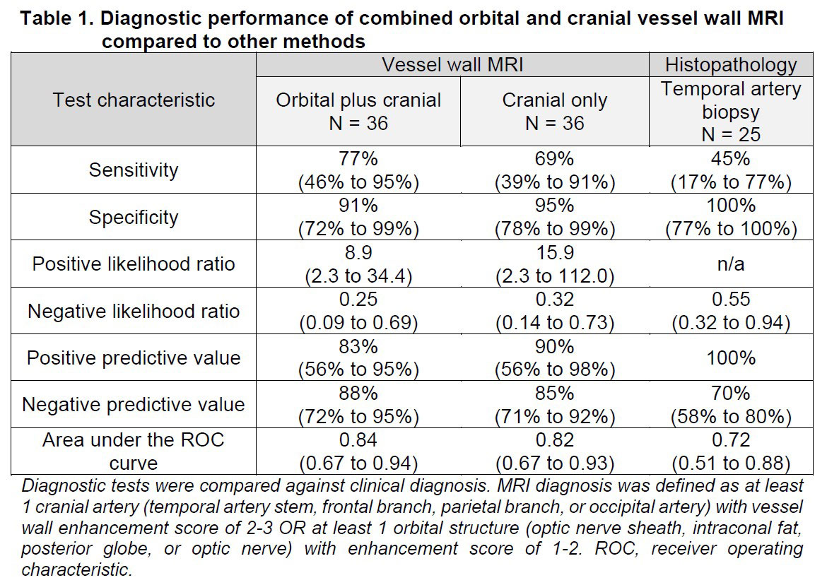
Methods: Patients with suspected new or relapsing cranial GCA who underwent orbital and brain vessel wall MRI were included. Nine patients had repeat imaging at months 1, 6, and 12 to assess changes over time. A clinical diagnosis of ocular or non-ocular GCA was determined by a rheumatologist and/or ophthalmologist and confirmed retrospectively. All MR images were scored by a single radiologist blinded to clinical data. Semi-quantitative integer scores of MRI enhancement were determined: 8 orbital structures were each scored 0, 1, or 2 (score 1-2 defined as abnormal) and included bilateral optic nerve sheath, optic nerve, intraconal orbital fat, and retrobulbar fat of posterior globe; 8 cranial arteries were each scored 0, 1, 2, or 3 (score 2-3 defined as abnormal) and included bilateral temporal arteries (stem, frontal, and parietal branches) and occipital arteries. MRI was considered diagnostic for GCA if at least 1 orbital or cranial artery had abnormal enhancement. Test characteristics were determined. Mixed-effects models were used to compare changes in scores over time between arteries that were abnormal vs normal at baseline.

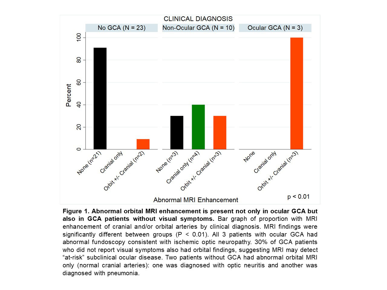
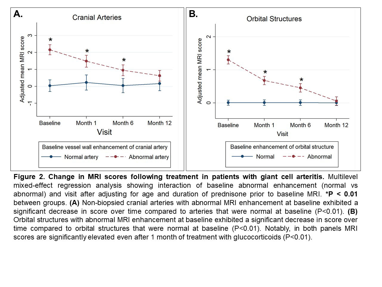
Results: The study included 36 patients with suspected cranial GCA: 31 new and 5 relapsing GCA. GCA was diagnosed in 13 patients: 10 non-ocular and 3 ocular GCA. The addition of orbital imaging to cranial vessel wall MRI had strong diagnostic performance with improved sensitivity compared to cranial MRI alone and temporal artery biopsy (Table 1). Orbital enhancement on MRI was observed in 100% of patients with ocular GCA and in 30% of GCA patients without visual symptoms (Figure 1). During follow-up, non-biopsied cranial arteries with abnormal MRI enhancement at baseline had a decrease in MRI score over time (P < 0.01, Figure 2A). Similarly, orbital MRI scores significantly decreased over time (P < 0.01, Figure 2B). Arteries with abnormal enhancement at baseline continued to have persistently elevated MRI scores even after 1 month of treatment (P< 0.01).

Conclusion: Combined orbital with cranial vessel wall MRI has very good diagnostic accuracy for GCA and may have the added value of identifying ocular GCA. This study is the first to demonstrate orbital MRI abnormalities in GCA patients without visual symptoms, suggesting that orbital MRI detects “at-risk” subclinical ocular disease. Imaging abnormalities persist even after 1 month of treatment, indicating flexibility in timing of MRI and potential translation to real-world settings. Decrease in MRI enhancement after treatment supports the possibility of using MRI for longitudinal disease surveillance. Future studies may determine if MRI identifies low-risk groups (e.g., those with normal orbital MRI) who can initiate a lower dose of glucocorticoids.

Supporting image 1

Supporting image 2

Supporting image 3


Disclosures: R. Rhee, None; S. Banerjee, None; V. Bhatt, None; M. Tamhankar, None; Q. Cao, None; N. Amudala, None; S. Chou, None; M. Burke, None; L. Loevner, Guerbet; P. Merkel, AbbVie, AstraZeneca, Boeringher-Ingelheim, Bristol-Myers Squibb, ChemoCentryx, Forbius, Genentech/Roche, Genzyme/Sanofi, GlaxoSmithKline, InflaRx, Neutrolis, Takeda, CSL Behring, Dynacure, EMDSerono, Immagene, Jannsen, Kiniksa, Magenta, Novartis, Pfizer, Q32, Regeneron, Sparrow, Eicos, Electra, Kyverna, UpToDate; J. Song, None.

To cite this abstract in AMA style:

Rhee R, Banerjee S, Bhatt V, Tamhankar M, Cao Q, Amudala N, Chou S, Burke M, Loevner L, Merkel P, Song J. Combined Orbital and Cranial Vessel Wall MRI for Diagnosis and Longitudinal Monitoring of Giant Cell Arteritis [abstract]. Arthritis Rheumatol. 2022; 74 (suppl 9). https://acrabstracts.org/abstract/combined-orbital-and-cranial-vessel-wall-mri-for-diagnosis-and-longitudinal-monitoring-of-giant-cell-arteritis/. Accessed .
  • Tweet
  • Click to email a link to a friend (Opens in new window) Email
  • Click to print (Opens in new window) Print

« Back to ACR Convergence 2022

ACR Meeting Abstracts - https://acrabstracts.org/abstract/combined-orbital-and-cranial-vessel-wall-mri-for-diagnosis-and-longitudinal-monitoring-of-giant-cell-arteritis/

Advanced Search

Your Favorites

You can save and print a list of your favorite abstracts during your browser session by clicking the “Favorite” button at the bottom of any abstract. View your favorites »

All abstracts accepted to ACR Convergence are under media embargo once the ACR has notified presenters of their abstract’s acceptance. They may be presented at other meetings or published as manuscripts after this time but should not be discussed in non-scholarly venues or outlets. The following embargo policies are strictly enforced by the ACR.

Accepted abstracts are made available to the public online in advance of the meeting and are published in a special online supplement of our scientific journal, Arthritis & Rheumatology. Information contained in those abstracts may not be released until the abstracts appear online. In an exception to the media embargo, academic institutions, private organizations, and companies with products whose value may be influenced by information contained in an abstract may issue a press release to coincide with the availability of an ACR abstract on the ACR website. However, the ACR continues to require that information that goes beyond that contained in the abstract (e.g., discussion of the abstract done as part of editorial news coverage) is under media embargo until 10:00 AM ET on November 14, 2024. Journalists with access to embargoed information cannot release articles or editorial news coverage before this time. Editorial news coverage is considered original articles/videos developed by employed journalists to report facts, commentary, and subject matter expert quotes in a narrative form using a variety of sources (e.g., research, announcements, press releases, events, etc.).

Violation of this policy may result in the abstract being withdrawn from the meeting and other measures deemed appropriate. Authors are responsible for notifying colleagues, institutions, communications firms, and all other stakeholders related to the development or promotion of the abstract about this policy. If you have questions about the ACR abstract embargo policy, please contact ACR abstracts staff at [email protected].

Wiley

  • Online Journal
  • Privacy Policy
  • Permissions Policies
  • Cookie Preferences

© Copyright 2025 American College of Rheumatology