Session Information
Date: Sunday, November 10, 2019
Title: T Cell Biology & Targets in Autoimmune & Inflammatory Disease Poster
Session Type: Poster Session (Sunday)
Session Time: 9:00AM-11:00AM
Background/Purpose: Approximately 40% of SLE patients experience neurological and/or psychiatric disorders, including cognitive abnormalities, mood disorders, confusion, psychosis, and seizures. However, the pathogenesis of neuropsychiatric SLE (NPSLE) is complex, and as of yet incompletely understood. We have previously demonstrated that the brain choroid plexus (CP) in the spontaneous MRL/lpr lupus mouse model, a strain which displays affective abnormalities and cognitive dysfunction similar to human NPSLE, is heavily infiltrated with effector T cells. In this study, we investigated T cell receptor (TCR) usage and repertoire diversity in CP infiltrating T cells, to enhance our understanding of this T cell population and its possible contribution to neuropsychiatric disease. In addition, since MRL/lpr mice exhibits T cell proliferation in other parenchymal organs (e.g. lungs and salivary glands), we sought to confirm that the brain T cell infiltration is organ specific.
Methods: Choroid plexuses and spleens were dissected from female MRL/lpr mice (n=5 per group) and MRL/MpJ mice (n=5) (the congenic control) at 16 weeks of age, by which time MRL/lpr mice display profound neurobehavioral deficits. The TCR repertoire was evaluated by isolating genomic DNA from choroid plexus and spleen, and amplifying the CDR3 sequence by multiplex PCR (Adaptive Technologies). The samples were evaluated with the Adaptive Technologies immunoSeq Analyzer and the R package, LymphoSeq.
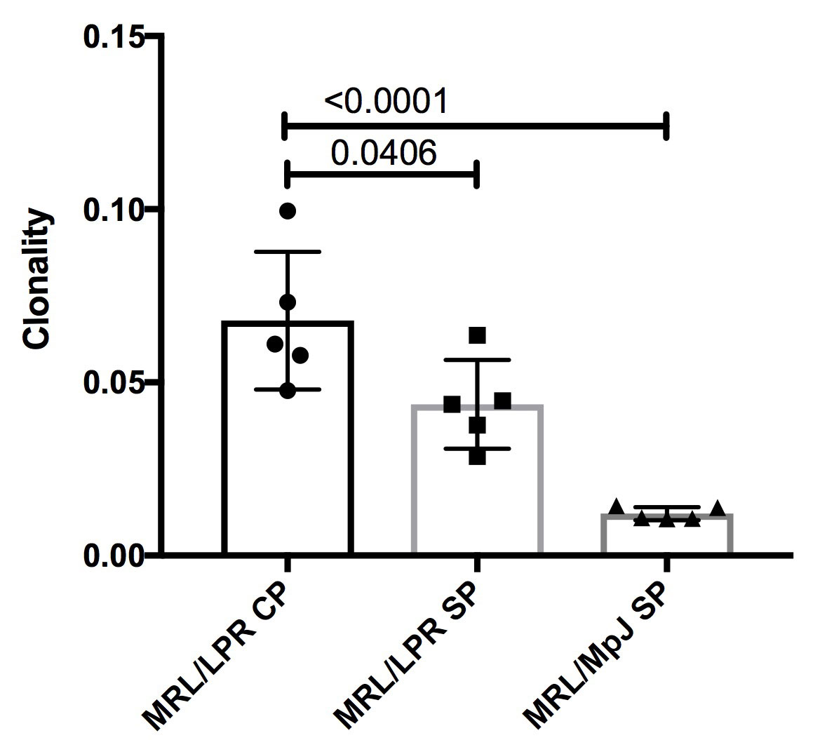
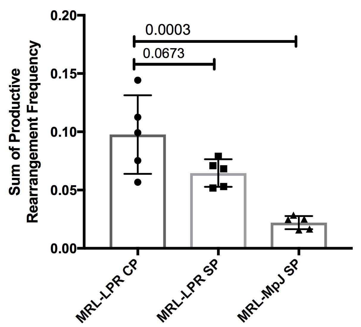
Results: MRL/lpr CP tissues had significantly increased clonality, derived from the Shannon entropy, as compared to both MRL/lpr and MRL/MpJ splenic tissues (p = 0.0406 and p < 0.0001 respectively). Similarly, T cells infiltrating the CP demonstrated enhanced oligoclonality, as seen by higher Gini coefficients (p = 0.0074 and p < 0.0001, respectively, Figure 1A). The abundance of the top 10 productive rearrangements was increased in MRL/lpr CP when compared to the MRL/lpr and MRL/MpJ splenic tissues (p = 0.0673 and p = 0.0003 respectively), as was the maximum productive frequency (Figure 1B). There was no difference in the gene usage or CDR3 length between the groups. Interestingly, there was an increased sample overlap for tissues originating from the same mouse rather than by organ type.
Conclusion: The results indicate that there is an increased oligoclonal expansion of T cells occurring in the CP of MRL/lpr mice compared to splenic tissues, suggesting antigen-driven brain T cell infiltration and expansion. Further comparative phenotyping of T cells found in different organs (lung, salivary glands, kidney) in the lupus strain is in progress.
To cite this abstract in AMA style:
Moore E, Huang M, Putterman C. Characterizing the Brain T Cell Receptor Repertoire in Neuropsychiatric Lupus [abstract]. Arthritis Rheumatol. 2019; 71 (suppl 10). https://acrabstracts.org/abstract/characterizing-the-brain-t-cell-receptor-repertoire-in-neuropsychiatric-lupus/. Accessed .« Back to 2019 ACR/ARP Annual Meeting
ACR Meeting Abstracts - https://acrabstracts.org/abstract/characterizing-the-brain-t-cell-receptor-repertoire-in-neuropsychiatric-lupus/


