ACR Meeting Abstracts

ACR Meeting Abstracts

  • Meetings
    • ACR Convergence 2024
    • ACR Convergence 2023
    • 2023 ACR/ARP PRSYM
    • ACR Convergence 2022
    • ACR Convergence 2021
    • ACR Convergence 2020
    • 2020 ACR/ARP PRSYM
    • 2019 ACR/ARP Annual Meeting
    • 2018-2009 Meetings
    • Download Abstracts
  • Keyword Index
  • Advanced Search
  • Your Favorites
    • Favorites
    • Login
    • View and print all favorites
    • Clear all your favorites
  • ACR Meetings

Abstract Number: 1035

Characterisation of New Bone Formation in Gout: A Quantitative Site-by-Site Analysis Using Plain Radiography and Computed Tomography

Nicola Dalbeth1, Aaron Milligan2, Barnaby Clark2, Fiona M. McQueen3 and Anthony Doyle4, 1Medicine, University of Auckland, Auckland, New Zealand, 2Department of Radiology, Auckland District Health Board, Auckland, New Zealand, 3Molecular Medicine and Pathology, University of Auckland, Auckland, New Zealand, 4University of Auckland, Auckland, New Zealand

Meeting: 2012 ACR/ARHP Annual Meeting

Keywords: bone disease, gout and imaging techniques

  • Tweet
  • Click to email a link to a friend (Opens in new window) Email
  • Click to print (Opens in new window) Print
Session Information

Title: Imaging of Rheumatic Diseases: Magnetic Resonance Imaging, Computed Tomography and X-ray

Session Type: Abstract Submissions (ACR)

Background/Purpose:   Radiographic descriptions of gout have noted the tendency to hypertrophic bone changes.  The aim of this study was to characterise the features of new bone formation (NBF) in gout, and to determine the relationship between NBF and other radiographic features of disease, particularly erosion and tophus. 

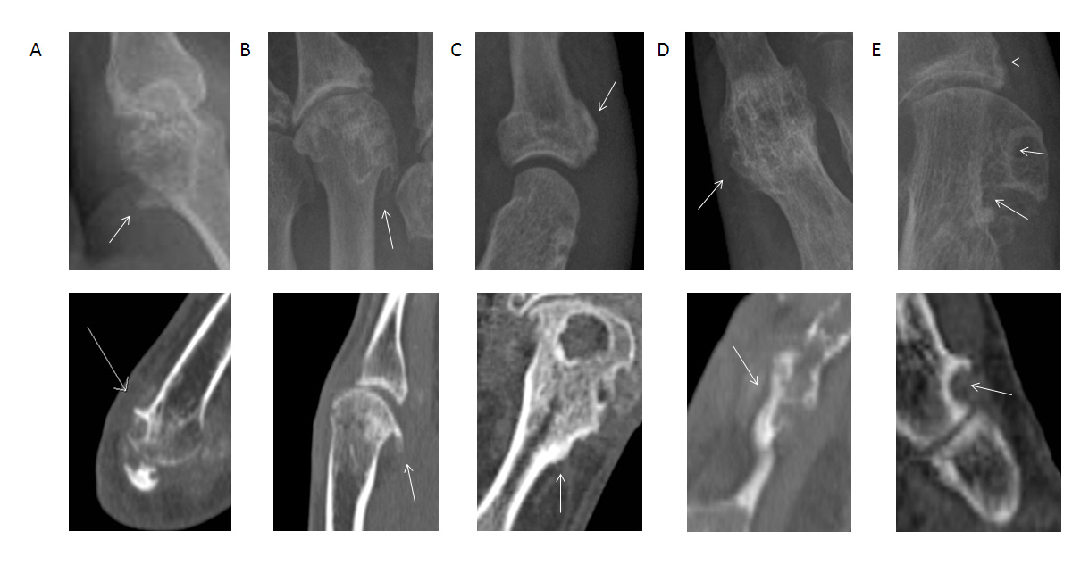
Methods: Paired plain radiographs (XR) and computed tomography (CT) scans of 798 individual hand and wrist joints from 20 patients with gout were analyzed.  Following a structured review of a separate set of images, films were scored for the presence of the following features of NBF: spur, osteophyte, periosteal NBF, ankylosis and sclerosis (Figure).  Sites for NBF scoring were those included in the gout-modified Sharp-van der Heijde method for erosion scoring. The relationship between NBF and other imaging features of gout (erosion and tophus) was analysed. 

Results: The most frequent forms of NBF were bone sclerosis (28.6% of all assessed joints using CT) and osteophyte (30.5%).  Spur and periosteal NBF were less common (17.8% and 6.0% respectively) , and ankylosis was rare (0.6%). On both XR and CT, joints with bone erosion were more likely to have NBF; for XR, odds ratios (OR) 64.9 for spur, 64.9 for osteophyte, 119.8 for periosteal NBF, 30.1 for ankylosis and 101.1 for sclerosis (p for all <0.0001); and for CT, OR 45.1 for spur, 3.3 for osteophyte, 16.6 for periosteal NBF, 26.6 for ankylosis and 32.3 for sclerosis (p for all <0.01).  Similarly, on CT, joints with intraosseous tophus were more likely to have NBF; OR 48.4 for spur, 3.3 for osteophyte, 14.5 for periosteal NBF, 35.1 for ankylosis and 39.1 for sclerosis (p for all <0.001). 

Conclusion: This detailed quantitative analysis has demonstrated that NBF occurs more frequently in joints affected by other features of gout.  This work suggests a connection between bone loss, tophus, and formation of new bone during the process of joint remodelling in gout. 

Figure.  Examples of the features of new bone formation (NBF) observed by plain radiography and computed tomography in patients with gout.  A. Spur. B. Osteophyte. C. Periosteal NBF. D. Ankylosis. E. Sclerosis.  Top panel shows plain radiographic images and lower panel shows CT images.


Disclosure:

N. Dalbeth,
None;

A. Milligan,
None;

B. Clark,
None;

F. M. McQueen,
None;

A. Doyle,
None.

  • Tweet
  • Click to email a link to a friend (Opens in new window) Email
  • Click to print (Opens in new window) Print

« Back to 2012 ACR/ARHP Annual Meeting

ACR Meeting Abstracts - https://acrabstracts.org/abstract/characterisation-of-new-bone-formation-in-gout-a-quantitative-site-by-site-analysis-using-plain-radiography-and-computed-tomography/

Advanced Search

Your Favorites

You can save and print a list of your favorite abstracts during your browser session by clicking the “Favorite” button at the bottom of any abstract. View your favorites »

All abstracts accepted to ACR Convergence are under media embargo once the ACR has notified presenters of their abstract’s acceptance. They may be presented at other meetings or published as manuscripts after this time but should not be discussed in non-scholarly venues or outlets. The following embargo policies are strictly enforced by the ACR.

Accepted abstracts are made available to the public online in advance of the meeting and are published in a special online supplement of our scientific journal, Arthritis & Rheumatology. Information contained in those abstracts may not be released until the abstracts appear online. In an exception to the media embargo, academic institutions, private organizations, and companies with products whose value may be influenced by information contained in an abstract may issue a press release to coincide with the availability of an ACR abstract on the ACR website. However, the ACR continues to require that information that goes beyond that contained in the abstract (e.g., discussion of the abstract done as part of editorial news coverage) is under media embargo until 10:00 AM ET on November 14, 2024. Journalists with access to embargoed information cannot release articles or editorial news coverage before this time. Editorial news coverage is considered original articles/videos developed by employed journalists to report facts, commentary, and subject matter expert quotes in a narrative form using a variety of sources (e.g., research, announcements, press releases, events, etc.).

Violation of this policy may result in the abstract being withdrawn from the meeting and other measures deemed appropriate. Authors are responsible for notifying colleagues, institutions, communications firms, and all other stakeholders related to the development or promotion of the abstract about this policy. If you have questions about the ACR abstract embargo policy, please contact ACR abstracts staff at [email protected].

Wiley

  • Online Journal
  • Privacy Policy
  • Permissions Policies
  • Cookie Preferences

© Copyright 2025 American College of Rheumatology