Session Information
Session Type: Poster Session (Sunday)
Session Time: 9:00AM-11:00AM
Background/Purpose: Carotid subclinical atherosclerosis is prevalent in patients with gout, although poorly predicted by cardiovascular risk assessment tools. Gout itself is deemed to contribute to its development. However, a previous report did not show association between clinical characteristics of gout and the presence of subclinical atherosclerosis [Ann Rheum Dis. 76:1263]. The aim of this study was to explore the association between sonographic signs of urate crystal deposits and carotid atherosclerosis.
Methods: Consecutive new patients with crystal-proven gout attended in a tertiary Rheumatology unit were eligible for the study. This included musculoskeletal and carotid ultrasound assessment, performed by a trained sonographer blinded to clinical data. Patients were examined during intercritical periods; flare prophylaxis with low-dose colchicine or other agents was permitted, but patients under urate-lowering treatment were excluded. The musculoskeletal scans evaluated wrists, 2nd MCPs and 1st MTPs joints, and triceps and patellar tendons, for the presence of signs suggestive of urate crystal deposits (double contour, hyperechoic aggregates, and tophi), following OMERACT definitions. Also, local power-Doppler (PD) signal was registered and graded as 0 to 3. The sum of locations showing crystal deposits or positive PD signal (≥1) was chosen to assess the crystal and inflammatory burden, respectively. Carotid arteries were scanned for increased intima-media thickness (IMT) and presence of atheroma plaques, according to Mannheim consensus. The association analysis was done by logistic regression, considering increased IMT or atheroma plaques as the independent variable.
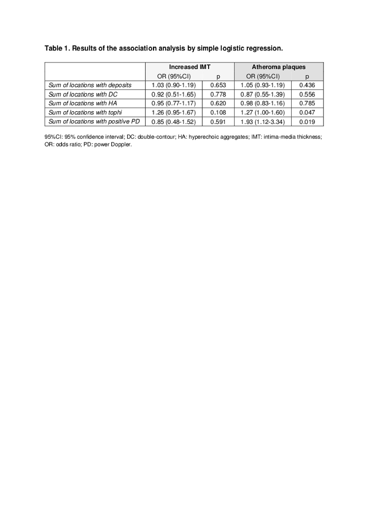
Results: Seventy-four new patients with gout were enrolled, mean aged 62.2 years (SD 14.7), 89.2% males. Mean gout duration was 5.6 years (SD 9.2), clinical tophi were observed in 15.9% of patients and mean serum urate level at diagnosis was 8.2 mg/dl (SD 1.6). All participants showed at least one sonographic sign of crystal deposits at the examined locations, with a mean sum of locations of 9 (SD 3.9). Regarding individual signs, their mean (SD) sum was as follows: 4.4 (2.2) for tophi, 3.7 (2.8) for aggregates and 0.8 (1.0) for double contour. The mean sum of locations with positive PD signal was 1.1 (SD 1.0). Regarding carotid scans, increased IMT was seen in 17 patients (24.3%), and atheroma plaques in 41 (55.4%). Table 1 shows the results of the association analysis. Tophi and positive PD signal were significantly associated with the presence of atheroma plaques, while no sonographic sign showed association with an increased IMT.
Conclusion: Sonographic deposits were consistently observed in new patients with gout. Tophi and positive PD signal, indicators of crystal and inflammatory burden, were significantly associated with carotid atheroma plaques. This new finding may contribute to understand the complex relationship between gout and atherosclerosis.
To cite this abstract in AMA style:
Calabuig I, Martínez-Sanchis A, Andrés M. Carotid Atherosclerosis and Sonographic Signs of Urate Crystal Deposits in Patients with Gout: An Association Study [abstract]. Arthritis Rheumatol. 2019; 71 (suppl 10). https://acrabstracts.org/abstract/carotid-atherosclerosis-and-sonographic-signs-of-urate-crystal-deposits-in-patients-with-gout-an-association-study/. Accessed .« Back to 2019 ACR/ARP Annual Meeting
ACR Meeting Abstracts - https://acrabstracts.org/abstract/carotid-atherosclerosis-and-sonographic-signs-of-urate-crystal-deposits-in-patients-with-gout-an-association-study/

