Session Information
Session Type: Abstract Submissions (ACR)
Background/Purpose
Data suggest that cerebral atrophy occurs in patients with SLE as early as within 9 months after disease onset. We aimed to address (1) if cerebral atrophy occurs even earlier after disease onset and (2) if glucocorticoid use early in the course of SLE would positively impact the brain gray matter (GMV) and white matter volumes (WMV).
Methods
Whole brain analyses of T1-weighted magnetic resonance (MR) images (1.5-T Siemen scanner) obtained ≤ 3 months of diagnosis and subsequently after sufficient disease control (SLEDAI<4) from 14 patients with new onset SLE (1997 ACR classification criteria) without clinically overt neuropsychiatric symptoms, traditional cardiovascular risk factors and antiphospholipid antibodies were performed. MR images of 14 demographically and intelligent-quotient matched healthy controls (HC) were used for comparison. An optimized voxel-based morphometry protocol was used to analyse the images. With thresholding at p<0.01 family-wise error (FWE) corrected, GMV and WMV between patients at diagnosis and HC were compared by 2-sample t-tests on 2-subject level probabilistic maps, while changes of GMV and WMV within the SLE group at diagnosis and after achieving sufficient disease control were compared by paired t-tests. Cumulative glucocorticoid doses were correlated with the changes of GMV and WMV of the regions of interest (ROI) within the SLE group.
Results
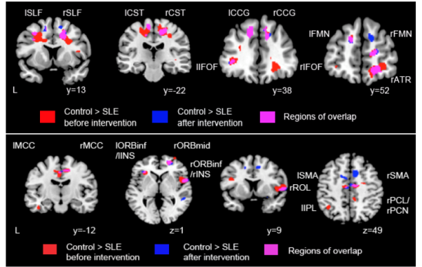
The mean±SD age of the patients at diagnosis and HC were 39.38±13.9 and 34.07±14.4 years respectively. The mean±SD SLEDAI and daily prednisolone dose were 9.93±5.7 and 15.32±18.4mg respectively. The mean±SD interval between diagnosis and the first MR scan was 36.86±35.5 days. Second MR scans in lupus patients were performed after a mean±SD of 504.92+267.0 days of treatment, with significant improvement of mean±SD SLEDAI and daily prednisolone dose to 2.64±1.4 (p<0.001) and 3.82±3.5mg (p=0.023) respectively. The mean±SD cumulative prednisolone was 1.76±2.10gm. Demonstrated in the first scans, lupus patients had significant GMV losses in both middle cingulate cortices and middle frontal gyrus, right rolandic operculum and the right supplementary motor area (lower panel of figure), and significant WMV losses in both superior longitudinal fasciculus, corticospinal tract, cingulum cingulate gyrus and inferior fronto-occipital fasciculus (upper panel of figure) as compared with HC. On ROI analyses, cumulative glucocorticoid dose was significantly correlated with improvement of brain volumes in the left inferior temporal gyrus, left cerebellum, and both anterior thalamic radiation in the SLE patients (all p<0.05).
Conclusion
In SLE patients, significant gray and white matter losses occurred as early as within a mean of 36.9 days of diagnosis. Glucocorticoid use early in the disease course improved GMV and WMV in certain areas, suggesting the potential benefit of glucocorticoids in brain plasticity and connectivity in early SLE.
Disclosure:
A. Mak,
None;
R. C. Ho,
None;
H. Tng,
None;
J. Zhou,
None.
« Back to 2014 ACR/ARHP Annual Meeting
ACR Meeting Abstracts - https://acrabstracts.org/abstract/brain-gray-and-white-matter-volume-losses-and-their-associations-with-glucocorticoid-use-in-patients-with-newly-diagnosed-systemic-lupus-erythematosus-sle-a-prospective-mr-study/

