ACR Meeting Abstracts

ACR Meeting Abstracts

  • Meetings
    • ACR Convergence 2024
    • ACR Convergence 2023
    • 2023 ACR/ARP PRSYM
    • ACR Convergence 2022
    • ACR Convergence 2021
    • ACR Convergence 2020
    • 2020 ACR/ARP PRSYM
    • 2019 ACR/ARP Annual Meeting
    • 2018-2009 Meetings
    • Download Abstracts
  • Keyword Index
  • Advanced Search
  • Your Favorites
    • Favorites
    • Login
    • View and print all favorites
    • Clear all your favorites
  • ACR Meetings

Abstract Number: 2583

Bone Structure and Perfusion Quantification of Bone Marrow Edema and Pannus Tissue Areas in the Wrist of Patients with RA

Jose R. Teruel Antolin1, Andrew J. Burghardt2, Julien Rivoire1, Waraporn Srikhum1, Susan M. Noworolski3, Thomas M. Link1, John B. Imboden4 and Xiaojuan Li5, 1Musculoskeletal Quantitative Imaging Research Group, Department of Radiology and Biomedical Imaging, University of California, San Francisco, San Francisco, CA, 2Department of Radiology & Biomedical Imaging, Musculoskeletal Quantitative Imaging Research, UCSF, San Francisco, CA, 3Radiology and Biomedical Imaging, University of California, San Francisco, San Francisco, CA, 4Department of Medicine, Division of Rheumatology, UCSF, San Francisco, CA, 5Department of Radiology and Biomedical Imaging, Musculoskeletal Quantitative Imaging Research, UCSF, San Francisco, CA

Meeting: 2012 ACR/ARHP Annual Meeting

Keywords: Bone density, Bone marrow, Computed tomography (CT), magnetic resonance imaging (MRI) and pannus

  • Tweet
  • Click to email a link to a friend (Opens in new window) Email
  • Click to print (Opens in new window) Print
Session Information

Title: Imaging of Rheumatic Diseases III: Computed Tomography

Session Type: Abstract Submissions (ACR)

Background/Purpose:

Bone marrow edema (BME) has been suggested as a strong predictor for erosive progression in RA joints, however, no previous studies examined the bone structure within BME. This study aimed to quantify bone structure and perfusion properties of BME, normal bone marrow (NBM) and pannus tissue areas in wrists affected by RA using 3T MRI and high resolution peripheral quantitative CT (HR-pQCT).

Methods:

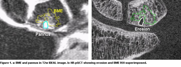
Sixteen patients (52.9 ± 12.7 years) fulfilling ACR classification were imaged in a HR-pQCT system (82µm isotropic voxel) and in a 3T MRI scanner using an 8-channel wrist coil. Coronal T2-weighted fast spin-echo IDEAL and 3D dynamic contrast enhanced (Gd-DTPA injection, temporal resolution 12 s, 32 time points) images were acquired. BME and pannus tissue areas were segmented semi-automatically in T2-w images. NBM areas were placed 1 to 3 mm far from BME regions and with similar distance to joint space. T2-w images were register to reformatted HR-pQCT and registration was applied to the segmented ROIs that were placed on original HR-pQCT images after upsampling (Fig 1). Perfusion parameters were calculated based on the signal-time curve obtained from DCE-MRI.

Figura3.JPG

Results:

Eleven out of 16 RA patients presented at least one BME region. Eighteen BME areas were segmented, 13 of them were presented around areas evidently affected by pannus tissue. The regions with pannus tissue in MRI always correspond to regions with erosion in HR-pQCT images (Fig 1). Two BME areas were presented next to early stage of pannus tissue (very small quantity of pannus penetrating the bone). For 3 of the BME regions, no pannus tissue were observed. Significant increases in bone density and trabecular thickness were evidenced in all BME regions (Fig 2).

figura2.JPG

BME and pannus tissue areas show significantly increased perfusion. The maximum signal enhancement in BME was 20.33 ± 14.33 (% over NBM) within the carpal bones and 21.32 ± 10.52 within the distal radius/ulna. In pannus tissue areas was 16.48 ± 11.34 (% over NBM) within the carpal bones and 32.93 ± 16.71 within the radius/ulna.

Conclusion:

BME regions show a thickening in the trabecular structure that suggests a bone regeneration before pannus tissue produces erosion. Trabecular thickening within BME is higher when pannus tissue is already presented in the patient. This higher thickening suggests sclerosis occurring to protect the bone structure from pannus tissue and kinematic re-adaptation. Combining MRI and HR-pQCT provides a powerful multi-modality approach for better understanding BME and erosion, and potentially identifying novel imaging markers for disease progression in RA.


Disclosure:

J. R. Teruel Antolin,
None;

A. J. Burghardt,
None;

J. Rivoire,
None;

W. Srikhum,
None;

S. M. Noworolski,
None;

T. M. Link,
None;

J. B. Imboden,
None;

X. Li,
None.

  • Tweet
  • Click to email a link to a friend (Opens in new window) Email
  • Click to print (Opens in new window) Print

« Back to 2012 ACR/ARHP Annual Meeting

ACR Meeting Abstracts - https://acrabstracts.org/abstract/bone-structure-and-perfusion-quantification-of-bone-marrow-edema-and-pannus-tissue-areas-in-the-wrist-of-patients-with-ra/

Advanced Search

Your Favorites

You can save and print a list of your favorite abstracts during your browser session by clicking the “Favorite” button at the bottom of any abstract. View your favorites »

All abstracts accepted to ACR Convergence are under media embargo once the ACR has notified presenters of their abstract’s acceptance. They may be presented at other meetings or published as manuscripts after this time but should not be discussed in non-scholarly venues or outlets. The following embargo policies are strictly enforced by the ACR.

Accepted abstracts are made available to the public online in advance of the meeting and are published in a special online supplement of our scientific journal, Arthritis & Rheumatology. Information contained in those abstracts may not be released until the abstracts appear online. In an exception to the media embargo, academic institutions, private organizations, and companies with products whose value may be influenced by information contained in an abstract may issue a press release to coincide with the availability of an ACR abstract on the ACR website. However, the ACR continues to require that information that goes beyond that contained in the abstract (e.g., discussion of the abstract done as part of editorial news coverage) is under media embargo until 10:00 AM ET on November 14, 2024. Journalists with access to embargoed information cannot release articles or editorial news coverage before this time. Editorial news coverage is considered original articles/videos developed by employed journalists to report facts, commentary, and subject matter expert quotes in a narrative form using a variety of sources (e.g., research, announcements, press releases, events, etc.).

Violation of this policy may result in the abstract being withdrawn from the meeting and other measures deemed appropriate. Authors are responsible for notifying colleagues, institutions, communications firms, and all other stakeholders related to the development or promotion of the abstract about this policy. If you have questions about the ACR abstract embargo policy, please contact ACR abstracts staff at [email protected].

Wiley

  • Online Journal
  • Privacy Policy
  • Permissions Policies
  • Cookie Preferences

© Copyright 2025 American College of Rheumatology