Session Information
Session Type: ACR Poster Session C
Session Time: 9:00AM-11:00AM
Background/Purpose: First degree relatives (FDR) of patients with RA are known to have approximately 4 times increased risk of developing RA and prevalence of RA is increased in Indigenous North Americans (INA) compared to Caucasian populations. To gain insight into immune phenotypes in those with increased risk of developing RA, we phenotyped B and T cell subsets in an INA cohort of FDR, RA patients and healthy controls (HC).
Methods: PBMC’s were isolated from FDR’s (n=10), RA patients (n=8) and HC (n=6) of INA ethnicity. B cell phenotyping (CD19, CD20, CD27, CD80, CD86, HLA-DR, Ki-67) and CD4 T cell phenotyping (CD3, CD4, CD45RA, CCR7, CXCR5, CXCR3, CCR2, PD-1, ICOS, Ki-67, HLA-DR) was performed by flow cytometry.
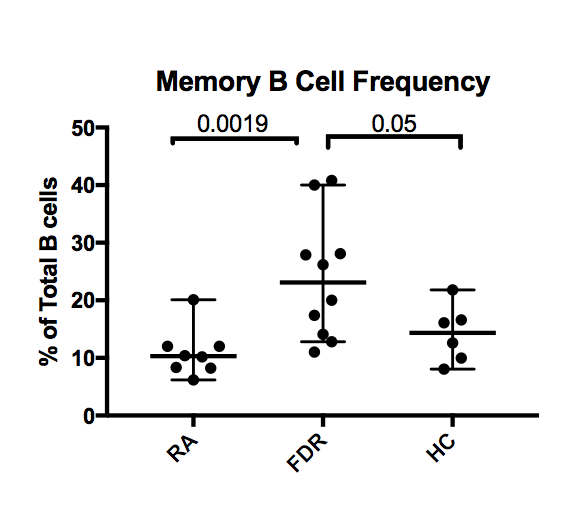
Results: FDR’s had increased frequency of CD20+27+ B cells (23.1% [IQR 13.8-31.1]) compared to RA patients (10.3% [8.3-12.0]) and seronegative controls (14.4% [9.5-17.9]) shown in Figure 1. The median fluorescence intensity (MFI) of CD27 in FDR was increased compared to HC (1393.5 [IQR 1268.1-1530.4] vs 1067.0 [880.5-2334.8]. There were no differences in the expression of CD80, CD86, HLA-DR or Ki-67 amongst memory (CD20+27+) and naïve (CD20+27-) B cell subsets.
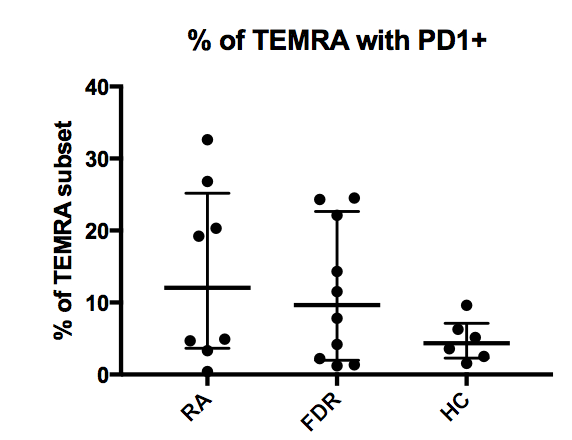
Distribution of CD4 T cell subsets (naïve, central memory, effector and terminal effector) between FDR, RA patients and HC was relatively similar. FDRs had double the frequency of PD-1 positive terminal effector memory (TEMRA) cells (9.7% [IQR 2.0-22.6]) compared to HC (4.4% [2.3-7.1]) shown in Figure 2. There was no difference in frequency of CXCR3, CCR2, Ki-67, ICOS or HLA-DR in these subsets amongst FDRs, HCs and RA patients.
Because CXCR5-/PD-1+ and CXCR5+/PD-1+ CD4 T cells are efficient in B cell stimulation, we also explored frequency of these T peripheral helper (Tph) and T follicular (Tfh) subsets amongst FDR, HC and RA patients. The Tph subset in the RA patients demonstrated increased ICOS (31.1% vs. 8.5%), Ki-67 (14.1% vs 5.5%), CCR2 (29.2% vs 22.0%), and HLA-DR (13.1% vs 8.0%) compared to FDR’s. All participants had similar frequencies of circulating Tfh and Tph subsets.
Conclusion: We found increased frequency of circulating memory B cells in the FDR population compared to unrelated controls. Whether these memory B cells are autoreactive in nature is not yet known. Increased PD-1 positive TEMRA cells in the FDR population, similar to the RA patients, suggests ongoing antigen experience and CD4 T cell activation in this group of people. Further characterization of regulatory mechanisms which prevent this high-risk group from developing autoimmune disease will be important in understanding the balance between pre-clinical autoimmunity and autoimmune disease.
Figure 1.
Figure 2.
To cite this abstract in AMA style:
Tanner S, Zhang C, Smolik I, Marshall A, El-Gabalawy H. B and T Cell Phenotypes of First Degree Relatives of RA Patients in an Indigenous North American Cohort [abstract]. Arthritis Rheumatol. 2018; 70 (suppl 9). https://acrabstracts.org/abstract/b-and-t-cell-phenotypes-of-first-degree-relatives-of-ra-patients-in-an-indigenous-north-american-cohort/. Accessed .« Back to 2018 ACR/ARHP Annual Meeting
ACR Meeting Abstracts - https://acrabstracts.org/abstract/b-and-t-cell-phenotypes-of-first-degree-relatives-of-ra-patients-in-an-indigenous-north-american-cohort/


