Session Information
Date: Monday, October 22, 2018
Title: Metabolic and Crystal Arthropathies – Basic and Clinical Science Poster I
Session Type: ACR Poster Session B
Session Time: 9:00AM-11:00AM
Background/Purpose: Lacking an immediate access to a polarized light microscope is sometimes used to justify the clinical diagnosis of crystal-related arthritis. Some studies have assessed the persistence of crystals in stored synovial fluid samples, with disparities in methodology and results that hamper the extraction of firm conclusions. The aim of this study was to determine the influence of the time from sampling and the method of preservation on the crystal identification in the synovial fluid samples under polarized light microscopy at different time points.
Methods: A prospective, longitudinal, observational study was designed. Synovial fluid samples were obtained in clinical practice (T0) and a trained rheumatologist identified crystals – monosodium urate (MSU) or calcium pyrophosphate (CPP). Fluids in which both types of crystals were detected, were excluded. On extraction, each fluid was divided into four samples. Two samples were stored in each type of tube – heparin or ethylenediaminetetraacetic acid (EDTA) as preserving agents -, at varying temperatures – room temperature or refrigerated at 4ºC (39.2ºF). Samples were analysed at around 24h (T1), 72h (T2), and 7 days (T3) by simple polarized light microscopy, and the presence of crystals was recorded. A medical student, who underwent training in crystal identification before the study, performed synovial fluid analyses; this training was repeated after every ten samples. The student was blinded to baseline data, and synovial fluid samples containing no crystals were included to ensure blinding. Differences in the proportion of crystal-containing samples throughout the time-points were assessed using Cochran’s Q and McNemar’s tests.
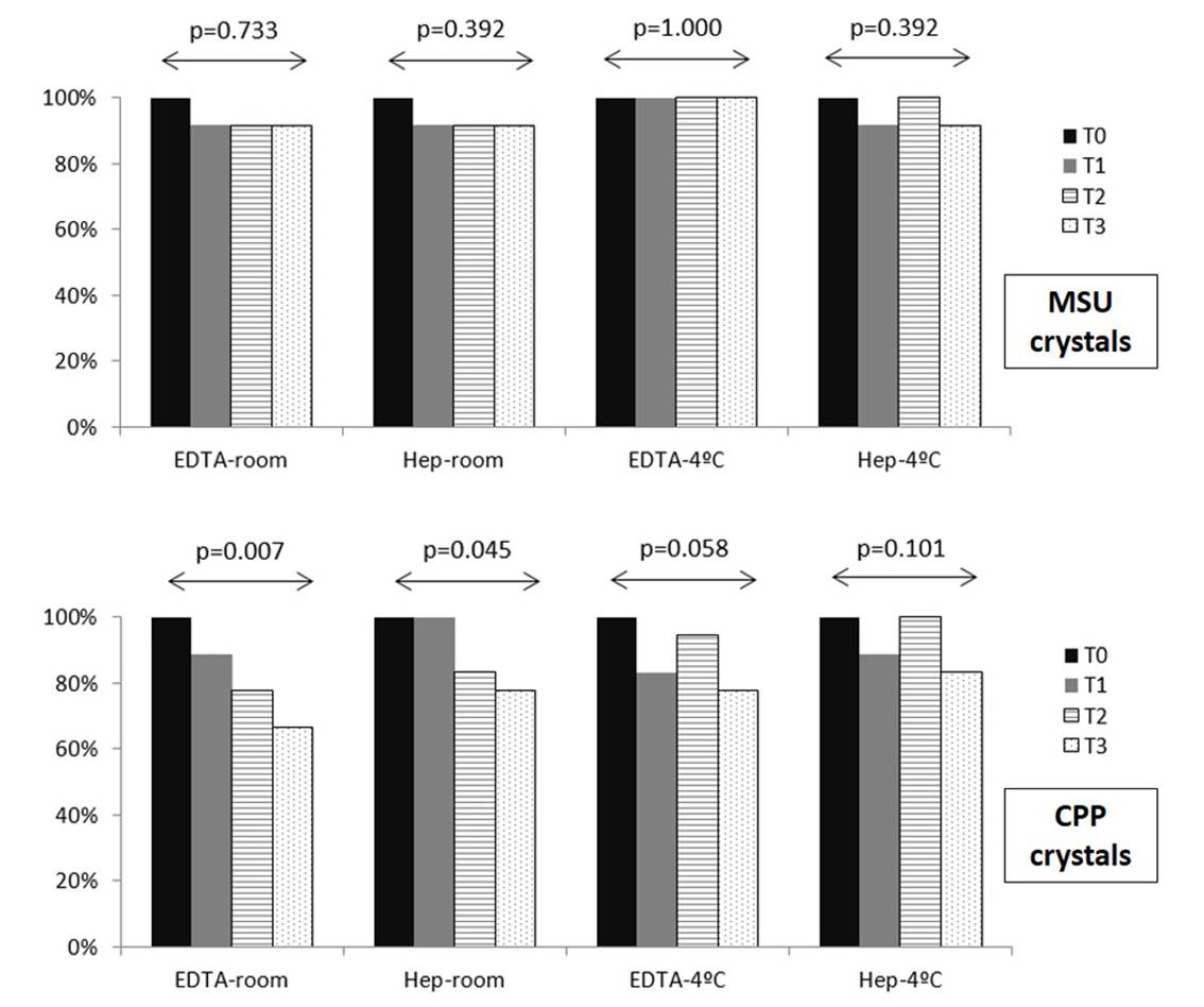
Results: Thirty synovial fluid samples were analysed, 12 with MSU and 18 with CPP crystals at baseline. This meant a total of 120 samples at follow-up time points. A total of 360 microscope visualizations were performed, at a mean (SD) of 31.0h (10.3) for T1, 90.5h (29.3) for T2, and 7.5days (0.7) for T3. The Figure shows the rate of samples containing crystals at different time-points. The identification of crystals within the MSU samples did not modify throughout study, being observed in 11/12 (91.7%) of room temperature samples and in 12/12 (100%) of refrigerated samples at T3. However, the identification of CPP crystals tended to decrease in all conditions, especially when preserved with EDTA and kept at room temperature (12/18 (66.7%) at T3).
Conclusion: MSU crystals persisted in stored samples for over a week, but CPP crystals tended to disappear in the same period, especially when kept at room temperature and/or preserved with EDTA. Keeping samples preserved with heparin and refrigerated at 4ºC appears to allow a delay microscope analysis for crystals. Foregoing crystal-proven diagnosis, due to immediate unavailability of a microscope, no longer appears justified.
To cite this abstract in AMA style:
Pastor S, Caño R, Gomez-Sabater S, Andrés M. Assessment of the Persistence of Crystals Under Polarised Light Microscopy in Stored Synovial Fluid Samples [abstract]. Arthritis Rheumatol. 2018; 70 (suppl 9). https://acrabstracts.org/abstract/assessment-of-the-persistence-of-crystals-under-polarised-light-microscopy-in-stored-synovial-fluid-samples/. Accessed .« Back to 2018 ACR/ARHP Annual Meeting
ACR Meeting Abstracts - https://acrabstracts.org/abstract/assessment-of-the-persistence-of-crystals-under-polarised-light-microscopy-in-stored-synovial-fluid-samples/

