Session Information
Session Type: Abstract Submissions (ACR)
Background/Purpose: 18F-fluoride uptake represents active osteoblastic bone synthesis. We explored structural damage of patients with ankylosing spondylitis (AS) using 18F-fluoride positron emission tomography (PET)-magnetic resonance imaging (MRI).
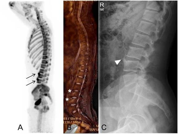
Methods: Whole spine of 6 male AS patients was examined with 18F-fluoride PET-MRI and conventional radiography. All participants were biologics naïve patients and fulfilled modified New York criteria. All images were assessed by two independent observers blinded for clinical data who recorded the presence or absence of increased 18F-fluoride uptake lesion in PET, acute (type A) and advanced (type B) corner inflammatory lesion (CIL) in MRI and syndesmophyte in conventional radiography at the anterior vertebral corners (Figure 1). Increased 18F-fluoride uptake was defined as an uptake which is greater than the uptake in the adjacent normal vertebral body. The association of increased 18F-fluoride uptake lesion with CIL and syndesmophyte was investigated by generalized linear latent mixed models analysis (GLLMM) to adjust within-patient dependence for total numbers of vertebral corners.
Results: There were 49 type A CIL (17.8%), 7 type B CIL (2.5%) and 38 increased 18F-fluoride uptake lesion (13.8%) out of 276 vertebral corners (C2 lower to SI upper) and 30 syndesmophyte (20.8%) out of 144 vertebral corners (C2 lower to T1 upper and T12 lower to SI upper). Increased 18F-fluoride uptake lesion was significantly associated with type A CIL (OR=6.9, 95% CI=3.1-15.2, p<0.001), type B CIL (OR=10.7, 95% CI=2.1-55.1, p=0.005) and syndesmpophyte (OR=55.6, 95% CI=7.3-422.3, p<0.001).
Conclusion: Our findings suggest that increased bone synthetic activity assessed by 18F-fluoride tracer uptake in the spine of AS patients can be associated with both inflammation and pre-existing new bone formation.
Figure 1. Forty-two-year-old male patient with ankylosing spondylitis. Increased 18F-fluoride uptake in positron emission tomography (arrows in A) and type B corner inflammatory lesion in short tau inversion recovery magnetic resonance image (asterisks in B) at L3 and 4 upper and corresponding syndesmophyte at L4 upper (arrowhead in C).
Disclosure:
G. Kim,
None;
S. G. Lee,
None;
S. H. Kim,
None;
J. W. Lee,
None;
S. H. Baek,
None.
« Back to 2013 ACR/ARHP Annual Meeting
ACR Meeting Abstracts - https://acrabstracts.org/abstract/assessment-of-structural-damage-in-patients-with-ankylosing-spondylitis-using-18f-fluoride-positron-emission-tomography-magnetic-resonance-imaging/

