ACR Meeting Abstracts

ACR Meeting Abstracts

  • Meetings
    • ACR Convergence 2024
    • ACR Convergence 2023
    • 2023 ACR/ARP PRSYM
    • ACR Convergence 2022
    • ACR Convergence 2021
    • ACR Convergence 2020
    • 2020 ACR/ARP PRSYM
    • 2019 ACR/ARP Annual Meeting
    • 2018-2009 Meetings
    • Download Abstracts
  • Keyword Index
  • Advanced Search
  • Your Favorites
    • Favorites
    • Login
    • View and print all favorites
    • Clear all your favorites
  • ACR Meetings

Abstract Number: 1645

Aortic Dilatation and PET/CT Vascular Activity at Diagnosis and 5 Years in an Inception Giant Cell Arteritis (GCA) Cohort

Anthony Sammel1, Ivan Ho Shon2, Daniel Moses1, Fredericks Stacey1, Gita Mathur1, Claudia Hillenbrand3, Edward Hsiao4, Schembri Geoffrey4, Rodger Laurent4 and Eva Wegner2, 1Prince of Wales Hospital, Randwick, Australia, 2The Prince of Wales Hospital, Sydney, Australia, 3University of NSW, Randwick, Australia, 4Royal North Shore Hospital, St. Leonards, Australia

Meeting: ACR Convergence 2023

Keywords: giant cell arteritis, Imaging

  • Tweet
  • Click to email a link to a friend (Opens in new window) Email
  • Click to print (Opens in new window) Print
Session Information

Date: Monday, November 13, 2023

Title: Abstracts: Vasculitis – Non-ANCA-Associated & Related Disorders I: Imaging

Session Type: Abstract Session

Session Time: 2:00PM-3:30PM

Background/Purpose: Aortic dilatation is typically a late complication of giant cell arteritis (GCA) but there are no tools to risk stratify patients at the time of diagnosis. We assessed the prevalence of aortic dilatation at 5 years in an inception giant cell arteritis cohort and assessed the relationship with FDG-PET/CT detected vascular activity at diagnosis, 6 months and 5 years.

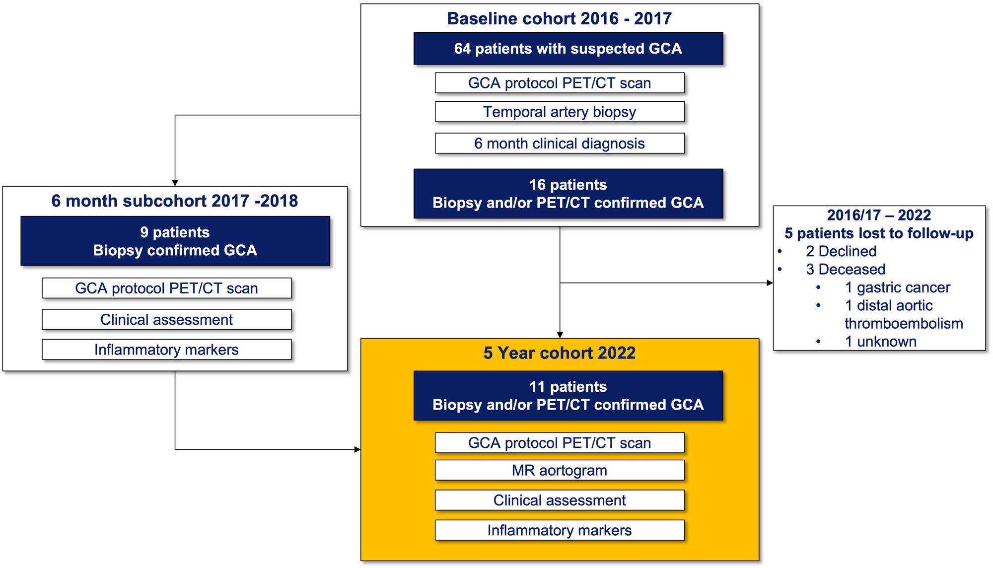
Methods: Patients were eligible for this 5-year study if they had been enrolled in the Giant Cell Arteritis and PET Scan (GAPS) cohort in 2016 or 2017, had a clinical diagnosis of GCA and a positive temporal artery biopsy (TAB) and/or a positive FDG-PET/CT scan at diagnosis. Patients underwent an FDG-PET/CT scan including assessment of cranial and large vessels, non-contrast MRI of the aorta, trans-thoracic echocardiogram (TTE) of the ascending aorta, blood collection and clinical assessment. PET/CT scans were dual reported by 2 blinded nuclear medicine physicians. Scans were reported overall positive or negative for disease activity and a visual grading of FDG avidity in each vascular territory was made with comparison to blood pool. MRI was reported by a single blinded cardiovascular radiologist and thoracic aortic dilatation was defined as external diameter >= 40 mm in ascending or >= 30 mm in the descending aorta.

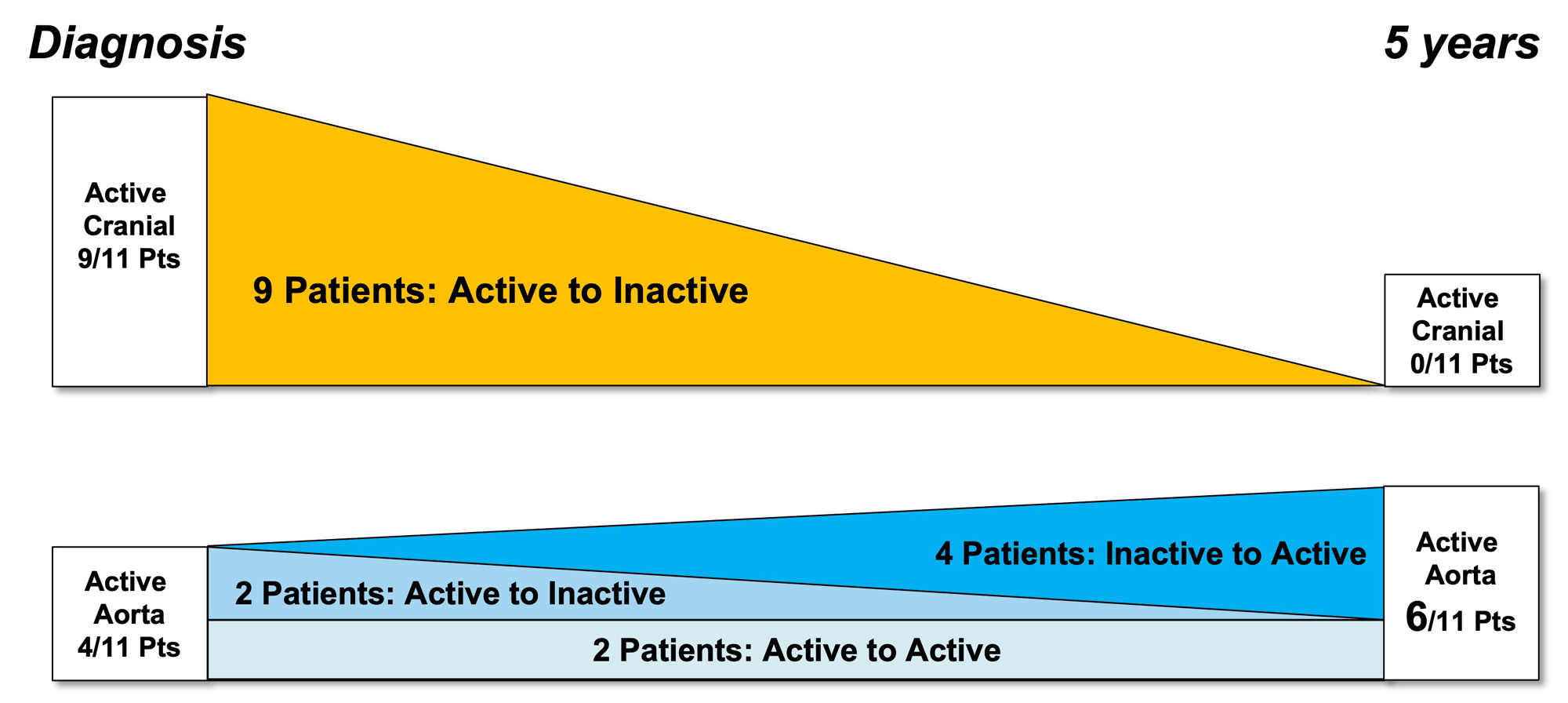
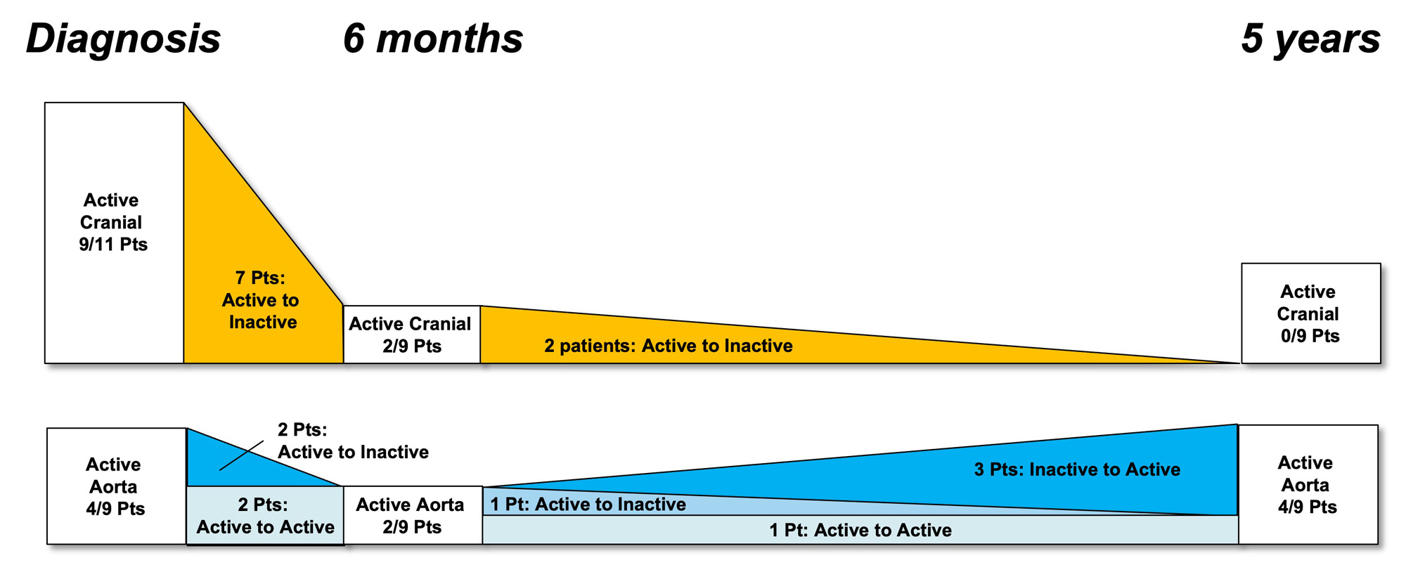
Results: 16 of the original 64 “suspected GCA” patients in the GAPS cohort met inclusion criteria and 11 participated in the 5-year study (3/16 deceased, 2/16 declined). The median age was 75, 73% were female and all were in clinical and serological remission with a median CRP of 1 (range 1 – 8). 4/11 (36%) patients had aortic dilatation (range 40 – 43mm) and 5/11 (45%) had globally active FDG-PET/CT scans. There was a trend towards a higher median aortic diameter in those with positive scans at 5 years (42 mm vs 35 mm, p=0.08) but aortic avidity at diagnosis did not predict 5-year dilatation. The distribution of FDG-PET/CT detected activity changed from a mix of cranial and large vessel disease at diagnosis to exclusively large vessel disease at 5-years. Aortitis developed in 4 patients who previously had inactive aortas. All 6 patients with inactive scans were taking an immunosuppressive agent (methotrexate, leflunomide, azathioprine or tocilizumab) at 5 years while all patients with active scans were not on therapy (p=0.02). TTE had excellent correlation with MRI for maximum aortic diameter (Pearson’s correlation coefficient 0.91).

Conclusion: Aortic dilatation was present in 36% GCA patients at 5 years. The distribution of FDG PET-CT detected avidity changed from predominantly cranial to exclusively large vessel disease and this may explain the lack of association between aortic avidity at diagnosis and dilatation at 5 years. Long-term use of steroid sparing agents may protect against subclinical vascular disease activity. TTE was a reliable screening modality for late aortic dilatation.

Supporting image 1

Figure 1: Patient flow

Supporting image 2

Figure 2: FDG-PET/CT activity by vascular region at diagnosis and 5 years

Supporting image 3

Figure 3: FDG-PET/CT activity by vascular region at diagnosis, 6 months and 5 years (subcohort who underwent 6 month scans)


Disclosures: A. Sammel: None; I. Ho Shon: None; D. Moses: None; F. Stacey: None; G. Mathur: None; C. Hillenbrand: None; E. Hsiao: None; S. Geoffrey: None; R. Laurent: None; E. Wegner: None.

To cite this abstract in AMA style:

Sammel A, Ho Shon I, Moses D, Stacey F, Mathur G, Hillenbrand C, Hsiao E, Geoffrey S, Laurent R, Wegner E. Aortic Dilatation and PET/CT Vascular Activity at Diagnosis and 5 Years in an Inception Giant Cell Arteritis (GCA) Cohort [abstract]. Arthritis Rheumatol. 2023; 75 (suppl 9). https://acrabstracts.org/abstract/aortic-dilatation-and-pet-ct-vascular-activity-at-diagnosis-and-5-years-in-an-inception-giant-cell-arteritis-gca-cohort/. Accessed .
  • Tweet
  • Click to email a link to a friend (Opens in new window) Email
  • Click to print (Opens in new window) Print

« Back to ACR Convergence 2023

ACR Meeting Abstracts - https://acrabstracts.org/abstract/aortic-dilatation-and-pet-ct-vascular-activity-at-diagnosis-and-5-years-in-an-inception-giant-cell-arteritis-gca-cohort/

Advanced Search

Your Favorites

You can save and print a list of your favorite abstracts during your browser session by clicking the “Favorite” button at the bottom of any abstract. View your favorites »

All abstracts accepted to ACR Convergence are under media embargo once the ACR has notified presenters of their abstract’s acceptance. They may be presented at other meetings or published as manuscripts after this time but should not be discussed in non-scholarly venues or outlets. The following embargo policies are strictly enforced by the ACR.

Accepted abstracts are made available to the public online in advance of the meeting and are published in a special online supplement of our scientific journal, Arthritis & Rheumatology. Information contained in those abstracts may not be released until the abstracts appear online. In an exception to the media embargo, academic institutions, private organizations, and companies with products whose value may be influenced by information contained in an abstract may issue a press release to coincide with the availability of an ACR abstract on the ACR website. However, the ACR continues to require that information that goes beyond that contained in the abstract (e.g., discussion of the abstract done as part of editorial news coverage) is under media embargo until 10:00 AM ET on November 14, 2024. Journalists with access to embargoed information cannot release articles or editorial news coverage before this time. Editorial news coverage is considered original articles/videos developed by employed journalists to report facts, commentary, and subject matter expert quotes in a narrative form using a variety of sources (e.g., research, announcements, press releases, events, etc.).

Violation of this policy may result in the abstract being withdrawn from the meeting and other measures deemed appropriate. Authors are responsible for notifying colleagues, institutions, communications firms, and all other stakeholders related to the development or promotion of the abstract about this policy. If you have questions about the ACR abstract embargo policy, please contact ACR abstracts staff at [email protected].

Wiley

  • Online Journal
  • Privacy Policy
  • Permissions Policies
  • Cookie Preferences

© Copyright 2025 American College of Rheumatology