Session Information
Session Type: ACR Concurrent Abstract Session
Session Time: 2:30PM-4:00PM
Background/Purpose :
Interferon (IFN) signaling is upregulated
in dermatomyositis (DM) skin, but the relationship to
classic histopathologic features is unknown.
Methods :
Thirty-nine skin biopsies from patients with dermatomyositis underwent gene expression analysis using Affymetrix arrays, as well as histologic analysis by a
blinded dermatopathologist. Biopsies were scored
for severity of several cardinal histopathologic
features, including basal vacuolar change (BV), dermal mucin
deposition, and perivascular inflammation. Type I and type
II IFN scores were generated based on the average relative expression of
selected genes previously
demonstrated to be upregulated by IFN-alpha and IFN-gamma, respectively.
Expression levels of IFN transcripts were measured by quantitative PCR. Univariate analysis was performed to evaluate the
association between IFN scores and IFN transcripts with key histopathologic
features, with subsequent multivariate models constructed to include
potentially confounding variables such as biopsy site, medication use, age, and
length of disease.
Results :
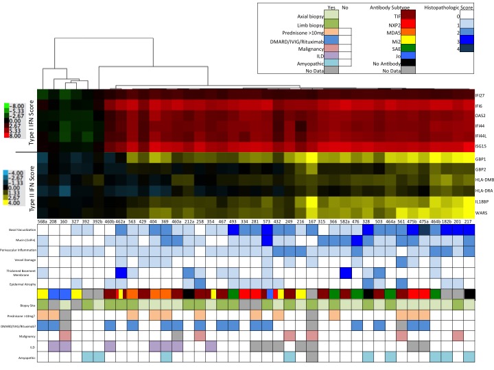
Skin biopsies were highly
heterogeneous regarding both histopathologic
features as well as type I or II IFN
scores. Type I IFN scores correlated strongly with
IFN beta expression (r=0.75,
p<0.0001) but not other
type I (alpha, kappa, omega) or type II IFN
transcript levels. In univariate
analysis, keratinocyte injury (reflected
by BV score) was found to be associated with both
type I and type II IFN scores (p=0.0014 and p=0.004, respectively); these associations
remained significant in multivariate analysis. In contrast, dermal
mucin deposition was highly correlated
with the type I IFN (p<0.0001) but not type II
IFN (p=0.071)
activity.
However, lymphocytic dermal
inflammation was associated with type II IFN score (p=0.0413), but
not the type I IFN score (p=0.3088). Additional
histologic features, including
epidermal atrophy, basement membrane thickening,
and vessel damage were not statistically associated with either
IFN score.
Conclusion :
Our results highlight that IFN signaling
in DM skin likely represents a combination of at least type I
and II IFN activity.
Furthermore,
our data suggest that type I and II signaling
are each associated with shared as well as distinct pathologic processes
in DM skin. Keratinocyte injury, the sine qua non finding
in DM skin, is associated
with both type I and II IFN activity. Finally, our
results imply that IFN beta is the
relevant IFN driving type I IFN signaling in DM skin and point
to both IFN beta and IFN gamma as potential targets of pharmacologic inhibition
in cutaneous DM.
To cite this abstract in AMA style:
Leatham HW, Rieger K, Chung L, Li S, Higgs BW, Yao Y, Sarin K, Fiorentino D. Activity of Type I and Type II Interferons in Dermatomyositis Skin Is Correlated with Characteristic Pathologic Features [abstract]. Arthritis Rheumatol. 2015; 67 (suppl 10). https://acrabstracts.org/abstract/activity-of-type-i-and-type-ii-interferons-in-dermatomyositis-skin-is-correlated-with-characteristic-pathologic-features/. Accessed .« Back to 2015 ACR/ARHP Annual Meeting
ACR Meeting Abstracts - https://acrabstracts.org/abstract/activity-of-type-i-and-type-ii-interferons-in-dermatomyositis-skin-is-correlated-with-characteristic-pathologic-features/

