Session Information
Date: Sunday, November 8, 2015
Title: Imaging of Rheumatic Diseases Poster I: Ultrasound, Optical Imaging and Capillaroscopy
Session Type: ACR Poster Session A
Session Time: 9:00AM-11:00AM
Background/Purpose:
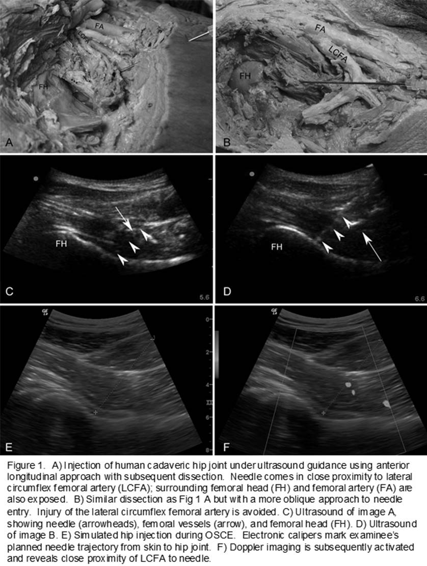
Injury of the lateral circumflex femoral artery (LCFA) is a potential cause of bleeding during invasive hip procedures due to its close proximity to the femoral acetabular joint. However, the LCFA is not routinely cited as a structure to avoid during intra-articular hip injections. The purpose of this study was to evaluate the risk of LCFA injury during ultrasound guided hip joint injection.
Methods:
1) We searched the PUBMED database (1967 to Apr 2015) for existing literature on basic techniques for ultrasound guided hip joint injection using the following search string: [“injection” OR “arthrocentesis” OR “aspiration”] AND [“hip” OR “intra-articular” OR “acetabulofemoral” OR “coxofemoral”] AND [“ultrasound” OR “ultrasonography” OR “ultrasound-guided” OR “image-guided”]. 52 non-English studies were excluded. 5 textbooks on musculoskeletal ultrasound were also reviewed. Resulting literature was screened for mention of the LCFA. 2) 18g spinal length needles were inserted under ultrasound guidance into the hip joints of 4 human cadavers. The tissues were dissected with the needles in place to expose the LCFA in relationship to the needle. 3) Rheumatologists trained for 8 months in ultrasound used electronic calipers to mark a planned needle trajectory from skin to hip joint on a live human ultrasound image during a musculoskeletal ultrasound final Observed Structured Clinical Examination (OSCE). Doppler imaging was subsequently used to locate the LCFA, and the closest distance between the planned needle trajectory and arterial signal was recorded.
Results:
1) 709 articles and 5 textbook chapters were reviewed; 11 discussed the technique of ultrasound guidance for hip injection. Of these 11 citations, 7 highlighted the femoral vascular bundle as a structure to avoid but only one specified routine use of Doppler imaging or specifically to avoid the LCFA. 2) In 3 out of the 4 human cadaveric dissections, the needle inserted into the hip under ultrasound guidance also made direct contact with the LFCA. 3) Of 27 OSCE participants, only 2 chose to activate Doppler imaging before marking their simulated hip injection trajectory. The electronic needle trajectory markings were subsequently found to pass through LFCA region Doppler signal in 6 (22%) cases. Among all 27 participants, the mean shortest distance from needle trajectory to arterial signal was 4mm (range 0-11mm).
Conclusion:
The risk of LCFA injury during ultrasound guided hip joint injection is substantial. Although the clinical significance of LCFA injury remains unclear, for high risk individuals (such as those on chronic anticoagulation), we suggest routine use of Doppler imaging as a part of standard hip injection protocols. Further study is required to determine if a more oblique or transverse approach to the hip will reduce the risk of accidental vascular injury.
To cite this abstract in AMA style:
Zhang M, Pessina M, Higgs JB, Kissin EY. A Vascular Obstacle in Ultrasound Guided Hip Joint Injection [abstract]. Arthritis Rheumatol. 2015; 67 (suppl 10). https://acrabstracts.org/abstract/a-vascular-obstacle-in-ultrasound-guided-hip-joint-injection/. Accessed .« Back to 2015 ACR/ARHP Annual Meeting
ACR Meeting Abstracts - https://acrabstracts.org/abstract/a-vascular-obstacle-in-ultrasound-guided-hip-joint-injection/