ACR Meeting Abstracts

ACR Meeting Abstracts

  • Meetings
    • ACR Convergence 2024
    • ACR Convergence 2023
    • 2023 ACR/ARP PRSYM
    • ACR Convergence 2022
    • ACR Convergence 2021
    • ACR Convergence 2020
    • 2020 ACR/ARP PRSYM
    • 2019 ACR/ARP Annual Meeting
    • 2018-2009 Meetings
    • Download Abstracts
  • Keyword Index
  • Advanced Search
  • Your Favorites
    • Favorites
    • Login
    • View and print all favorites
    • Clear all your favorites
  • ACR Meetings

Abstract Number: 2659

A Comprehensive Approach Utilizing a Combination of Genetics, Cytological Analysis, and Imaging Techniques to Speed up the Diagnostic Process in VEXAS Patients

Sara Bindoli1, Chiara Baggio2, Roberto Padoan3, Riccardo Bixio4, doria Andrea5, Roberta Ramonda2 and Paolo Sfriso1, 1Rheumatology Unit, University of Padova, Italy, Padova, Veneto, Italy, 2Rheumatology Unit, University of Padova, Italy, Padova, Italy, 3Department of Medicine DIMED, Division of Rheumatology, University of Padua, Padua, Italy, 4University of Verona, Verona, Verona, Italy, 5University of Padova, Padova, Italy

Meeting: ACR Convergence 2024

Keywords: Autoinflammatory diseases, genetics, Inflammation

  • Tweet
  • Click to email a link to a friend (Opens in new window) Email
  • Click to print (Opens in new window) Print
Session Information

Date: Tuesday, November 19, 2024

Title: Abstracts: Miscellaneous Rheumatic & Inflammatory Diseases II

Session Type: Abstract Session

Session Time: 11:00AM-12:30PM

Background/Purpose: VEXAS is a newly described “haemato-autoinflammatory” condition resulting from somatic mutations in the UBA-1 gene. By integrating genetics, cytology and imaging, this work seeks to advance a diagnostic pathway to better characterize VEXAS patients from the outset.

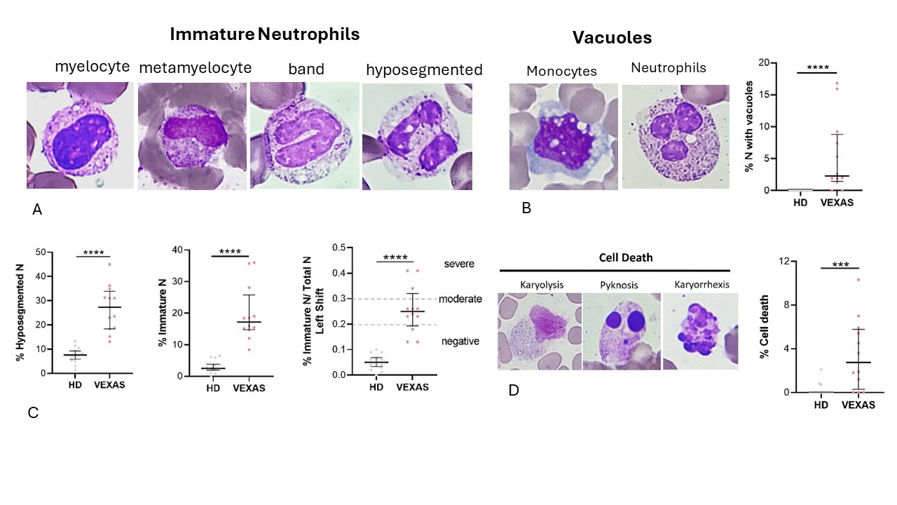
Methods: Fourteen consecutive males (median age 74.5, IQR 8.5) with suspected VEXAS syndrome and 16 healthy donors (HDs) were gathered. Whole blood from patients was collected for exome analysis (HiSeq2500 Illumina Sequencer) to assess the potential carriage of UBA-1 mutations. In parallel, peripheral blood cells were examined. May-Grünwald Giemsa staining was used to study the cellular morphology, including cytoplasm, granules, and vacuoles, and to perform a cytogenic evaluation of leukocytes. Interleukin (IL)-1β, TNFα, IL-1α, IL-18 and IL-8 were measured in plasma using commercially available enzyme-linked immunosorbent assay (ELISA) kits. As a preliminary work-up, positron-emission tomography/computed tomography (PET-CT) scans were executed; images were analysed drawing regions of interest (ROI), and a semi-quantitative analysis was performed for each scan by fixing the Standardized Uptake Values (SUVs).

Results: Twelve patients (86%) had mutations in the UBA-1 gene. Table 1 shows the characteristics of the patients with VEXAS. At cytological evaluation (Figure 1), hyposegmented and immature neutrophils (band cells, metamyelocytes, and myelocytes) were higher in VEXAS than HDs (p< 0.0001). A moderate shift to the left (0.2 – 0.29) and a severe shift to the left (≥ 0.3) were observed in 3 (25%) and 7 (58%) VEXAS patients, respectively; the I/T index was higher in VEXAS than HDs (p< 0.0001). Vacuoles were observed in monocytes and neutrophils of 10/12 (83%) patients and were absent in HDs (p< 0.0001). Although not significant, the percentage of neutrophils and immature neutrophils was higher in patients with the Met41Val mutation compared to the other variants. A significant difference was observed in the cell death rate between VEXAS and controls (p< 0.001). Regarding cytokines, no differences in the IL-1β levels were found between HDs and VEXAS. The levels of IL-18 (p< 0.0001), IL-1α (p< 0.05), TNFα (p< 0.01) and IL-8 (p< 0.05) were higher in VEXAS. Patients carrying the Met41Val variation had higher IL-1β, IL-18, IL-1α and IL-8 levels than those with other known mutations. The percentage of cell death correlated with IL-1β (r=0.840, p =0.001) and IL-8 (r=0.758, =0.006). Finally, IL-8 levels correlated with the vacuoles (r= 0.732, p=0.009) and micronuclei rates (r=0.777, p=0.005). PET-CT scans were performed in 7/12 (58%) patients showing diffuse hyper-uptakes mainly in bone marrow (57%) and lymph nodes (43%) (Table 1).

Conclusion: Cytological evaluation appears to be a rapid and non-invasive way of detecting abnormalities consistent with the suspected diagnosis while waiting for genetics, or as an alternative to bone marrow biopsy. To facilitate a prompt treatment start, the PET-CT scan may be helpful in ruling out mimicking pathologies such as solid cancer, lymphoproliferative diseases, and other poly/monogenic autoinflammatory conditions.

Supporting image 1

Table 1. Description of the clinical features, laboratory biomarkers and PET-CT findings of the VEXAS cohort; the risk for myelodysplastic syndrome was defined according to the R-IPSS (Revised International Prognostic Scoring System).
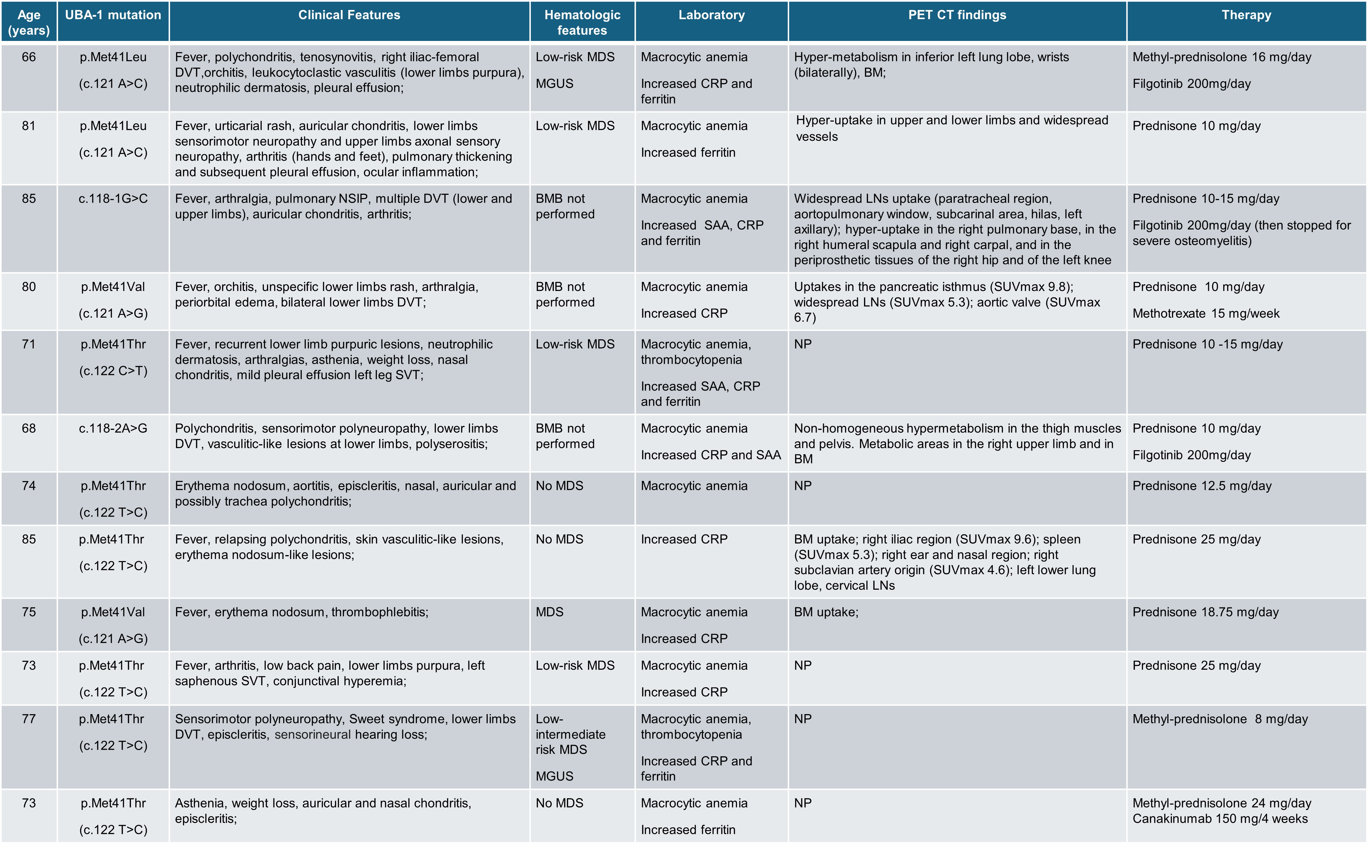
Legend: BM: bone marrow; BMB: bone marrow biopsy; CRP: C-reactive protein; DVT: deep vein thrombosis; LNs: lymphnodes; MDS: myelodysplastic syndrome; MGUS: monoclonal gammopathy of unknown significance; NP= not performed; NSIP: non-specific interstitial pneumonia; SAA: serum amyloid A; SUV: standardized uptake value; SVT: superficial vein thrombosis;

Supporting image 2

Figure 1. Cytogenic and cellular morphology evaluation of leukocytes in blood smear from VEXAS patients. Blood smears from VEXAS patients (n = 12) and HD (n = 16) were stained using MGG staining as described in Methods. (A) Representative image of immature and hyposegmented neutrophils stained with MGG. (B) Representative image of monocytes and neutrophils with vacuoles stained with MGG and vacuolization rates in neutrophils of VEXAS and HD. (C) Rate of hyposegmented, immature neutrophils and left shift in VEXAS patients and HD. (D) Representative image of karyorrhexis, karyolysis, and pyknosis in blood smears stained with MGG. Data are expressed as medians and IQR. p calculated using the Mann-Whitney test: ***p<0.001, ****p<0.0001.


Disclosures: S. Bindoli: None; C. Baggio: None; R. Padoan: None; R. Bixio: Accord Healthcare, 6, GlaxoSmithKlein(GSK), 6; d. Andrea: AstraZeneca, 2, 6, Bristol-Myers Squibb(BMS), 2, 6, Eli Lilly, 2, 6, Galapagos, 2, 6, GlaxoSmithKlein(GSK), 2, 6, Janssen, 2, 6, Otsuka, 2, 6; R. Ramonda: AbbVie/Abbott, 6, Eli Lilly, 2, 6, Novartis, 1, 6, Pfizer, 6, UCB, 6; P. Sfriso: None.

To cite this abstract in AMA style:

Bindoli S, Baggio C, Padoan R, Bixio R, Andrea d, Ramonda R, Sfriso P. A Comprehensive Approach Utilizing a Combination of Genetics, Cytological Analysis, and Imaging Techniques to Speed up the Diagnostic Process in VEXAS Patients [abstract]. Arthritis Rheumatol. 2024; 76 (suppl 9). https://acrabstracts.org/abstract/a-comprehensive-approach-utilizing-a-combination-of-genetics-cytological-analysis-and-imaging-techniques-to-speed-up-the-diagnostic-process-in-vexas-patients/. Accessed .
  • Tweet
  • Click to email a link to a friend (Opens in new window) Email
  • Click to print (Opens in new window) Print

« Back to ACR Convergence 2024

ACR Meeting Abstracts - https://acrabstracts.org/abstract/a-comprehensive-approach-utilizing-a-combination-of-genetics-cytological-analysis-and-imaging-techniques-to-speed-up-the-diagnostic-process-in-vexas-patients/

Advanced Search

Your Favorites

You can save and print a list of your favorite abstracts during your browser session by clicking the “Favorite” button at the bottom of any abstract. View your favorites »

All abstracts accepted to ACR Convergence are under media embargo once the ACR has notified presenters of their abstract’s acceptance. They may be presented at other meetings or published as manuscripts after this time but should not be discussed in non-scholarly venues or outlets. The following embargo policies are strictly enforced by the ACR.

Accepted abstracts are made available to the public online in advance of the meeting and are published in a special online supplement of our scientific journal, Arthritis & Rheumatology. Information contained in those abstracts may not be released until the abstracts appear online. In an exception to the media embargo, academic institutions, private organizations, and companies with products whose value may be influenced by information contained in an abstract may issue a press release to coincide with the availability of an ACR abstract on the ACR website. However, the ACR continues to require that information that goes beyond that contained in the abstract (e.g., discussion of the abstract done as part of editorial news coverage) is under media embargo until 10:00 AM ET on November 14, 2024. Journalists with access to embargoed information cannot release articles or editorial news coverage before this time. Editorial news coverage is considered original articles/videos developed by employed journalists to report facts, commentary, and subject matter expert quotes in a narrative form using a variety of sources (e.g., research, announcements, press releases, events, etc.).

Violation of this policy may result in the abstract being withdrawn from the meeting and other measures deemed appropriate. Authors are responsible for notifying colleagues, institutions, communications firms, and all other stakeholders related to the development or promotion of the abstract about this policy. If you have questions about the ACR abstract embargo policy, please contact ACR abstracts staff at [email protected].

Wiley

  • Online Journal
  • Privacy Policy
  • Permissions Policies
  • Cookie Preferences

© Copyright 2025 American College of Rheumatology