Session Information
Date: Sunday, November 8, 2015
Title: Imaging of Rheumatic Diseases Poster I: Ultrasound, Optical Imaging and Capillaroscopy
Session Type: ACR Poster Session A
Session Time: 9:00AM-11:00AM
Background/Purpose:
Even though Doppler ultrasound (US) is used for
diagnosing inflammation in arthritides, it is well-known that Doppler signals
may be seen in healthy wrist and finger joints (1). Tenosynovitis has been shown to be frequent in rheumatoid
arthritis and to predict erosive disease (2). Detailed knowledge of the distribution of feeding vessels in
fingers is important to distinguish normal from pathological findings. However,
there is no knowledge about Doppler signals in relation to healthy tendon
sheaths and the possible pitfalls this may generate.
To investigate presence of feeding vessels in relation
to the healthy flexor and extensor tendon sheaths of the wrist by use of 3D Doppler
US.
Methods:
Twenty healthy participants were recruited; 10 women
in the age 27-54 years and 10 men in the age 27-59 years. None of the
participants had finger pain, history of arthritis or any known finger tendon
disease, or were smokers.
The participants had US of the palmar and
dorsal side of the right wrist. US was carried out using a General Electric
Logiq E9 with a 3D ultrasound probe. The Doppler settings were adjusted
according to published recommendations (3) with a
Doppler frequency of 8.3 MHz and pulse repetition frequency of 0.4. The same
Doppler settings were used for all examinations. Specific probe positions on the
wrist were selected before study initiation at two different levels (Lister’s
tubercle and pisiforme). Two scans were made at each position to minimise the risk
of missing Doppler findings due to different parts of the cardiac cycle being
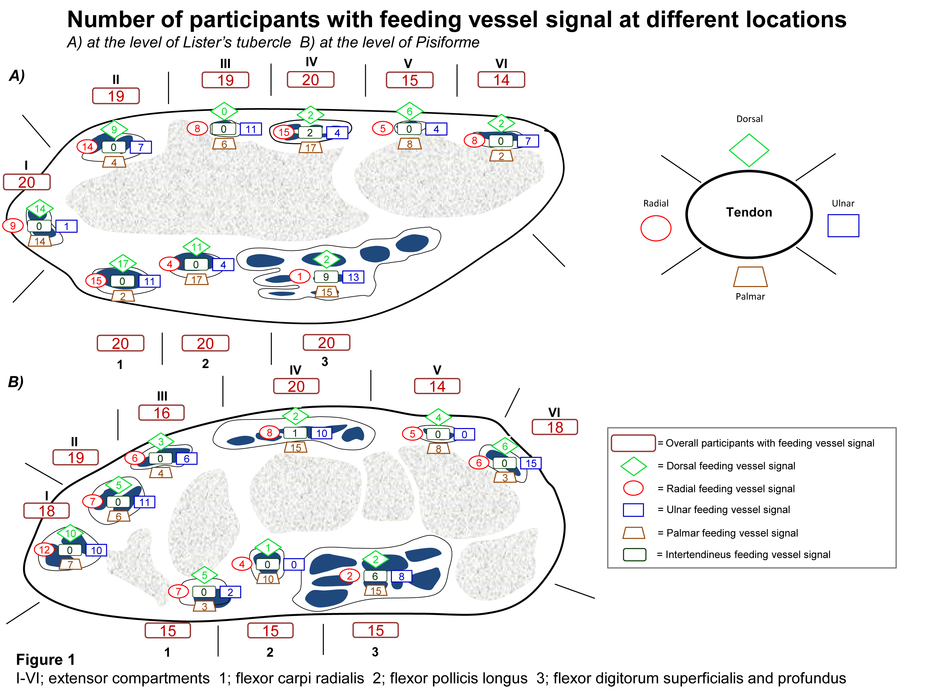
sampled as the sweep was made. Each tendon sheath was divided into specific
areas and the visualized 3D Doppler findings in relation to the tendon sheath
were plotted on a schematic drawing (Figure 1A and 1B).
Results:
The overall distribution of feeding vessels
was comparable at the level of Lister’s tubercle and the level at the pisiforme
for the extensor tendons. For the flexor tendons, feeding vessels were more
frequent at the level of Lister’s tubercle, as shown in figure 1(A and B). Overall
feeding vessels were less frequent for compartment V and VI at the level of
Lister’s tubercle. Feeding vessels were seen less at the superficial location
for the extensor tendons, except for compartment I. Radial and dorsal vessels
were rare in the tendon sheath of flexor digitorum superficialis and profundus.Intertendineus
feeding vessels were mainly seen in the tendon sheath of flexor digitorum
superficialis and profundus.
Conclusion:
Feeding vessels in close relation to the extensor and
flexor tendon sheaths were common in the wrist of healthy participants and may
be a cause of misinterpretation due to artefacts. These vessels should be taken
into consideration when diagnosing tenosynovitis in the wrist.
To cite this abstract in AMA style:
Ammitzbøll-Danielsen M, Janta I, Torp-Pedersen S, Naredo E, Østergaard M, Terslev L. 3D Ultrasound Doppler Findings in Wrist Tendon Sheaths of Healthy Controls [abstract]. Arthritis Rheumatol. 2015; 67 (suppl 10). https://acrabstracts.org/abstract/3d-ultrasound-doppler-findings-in-wrist-tendon-sheaths-of-healthy-controls/. Accessed .« Back to 2015 ACR/ARHP Annual Meeting
ACR Meeting Abstracts - https://acrabstracts.org/abstract/3d-ultrasound-doppler-findings-in-wrist-tendon-sheaths-of-healthy-controls/