Session Information
Session Type: Abstract Submissions (ACR)
Background/Purpose:
SLE patients have perturbations in B cell subsets including a large expansion of IgD-CD27- B cells (DN) in patients with active disease. Patients also have changes in PBMC gene expression, particularly increased expression of interferon regulated genes (IRG). However, data on gene expression in SLE B cells is limited. The purpose of this study was to understand the function and potential dysregulation of DN in SLE by comparing differences in gene transcription between B cell subsets from SLE patients and healthy control donors (HCD).
Methods:
RNA was isolated from B cells from 3 HCD and 3 SLE patients with elevated DN sorted into naive (IgD+CD27-CXCR5+), switched memory (IgD-CD27+CXCR5+), and DN (IgD-CD27-CXCR5-) subsets. After amplification, high throughput sequencing of cDNA was used for transcriptional expression profiling. Sequencing data was processed and analyzed using the Tuxedo suite and EdgeR software package. Genes were tested for differential expression using a three way ANOVA model.
Results:
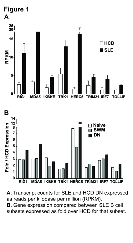
115 of the 249 genes differentially expressed in B cells between SLE patients and HCD were IRG. In addition to previously defined IRG, pathway analysis found several genes involved in TLR-3 and RNA sensing signaling pathways. These included RNA sensing molecules RIG1 and MDA5, downstream kinases IKBKE and TBK1, kinase substrate and transcription factor IRF-7, and HERC5 and TRIM21, molecules that regulate IRF stability. Of these only IRF-7 has been previously described as differentially expressed in SLE B cells. While, expression for most genes was increased in all B cell subsets, the DN subset showed the largest increase in expression for all genes except IKBKE and TRIM21. The DN subset also had increased expression and differential splicing of TOLLIP, an inhibitor of MyD88 mediated TLR signaling.
Conclusion:
IKBKE and TBK1 are key signaling kinases for detecting RNA through the TLR3 and MDA5/RIG1 pathways. These pathways detect viral RNA but are also activated by endogenous RNA from apoptotic cells. Increased expression of IKBKE and TBK1 in SLE B cells is accompanied by the expression of both the upstream receptors and downstream kinase substrate. Furthermore, while TRIM21 mediates degradation of IRF-7, expression of TRIM21 was lowest in the DN and these cells expressed high levels of HERC5, which prevents IRF degradation. Coordinated expression of these genes strongly suggests these pathways are more active in SLE B cells and particularly in the DN population. In SLE, RNA from apoptotic cells can be internalized in RNP and Ro60 immune complexes through Fc receptors(FCR) and are thus available to MDA5/RIG1 and TLR-3. DN express high levels of FCR. This combined with increased expression of these signaling molecules likely results in RNA antigens having a powerful pro-inflammatory influence on DN.
Disclosure:
S. Jenks,
None;
E. Ramos,
None;
I. Sanz,
Pfizer Inc ,
5,
Biogen Idec,
9.
« Back to 2013 ACR/ARHP Annual Meeting
ACR Meeting Abstracts - https://acrabstracts.org/abstract/igd-cd27-b-cells-from-systemic-lupus-erythematous-patients-have-increased-expression-of-genes-involved-in-rna-sensing-and-toll-like-receptor-3-signaling-pathways/

