Session Information
Session Type: Abstract Submissions (ACR)
Background/Purpose: Osteoarthritis is a degenerative disease without a treatment. Tissue Engineering could provide an alternative tool for cartilage repair. The aim of this study was to evaluate the neotissue formed using human bone marrow mesenchymal stem cells (hBM-MSCs) and collagen scaffolds.
Methods: hBM-MSCs were cultured on collagen (Col), Col+heparan sulfate (HS), Col+chondroitin sulfate (CHS) and Col+heparin (OLH3) scaffolds, in chondrogenic medium supplemented by TGFβ-3. Chondrogenic differentiation and the constructs were evaluated by histochemistry, immunohistochemistry, and molecular biology. Cellular morphology and ultrastructure were studied by electron microscopy. Culture supernatants were collected and the amount of secreted collagen was measured by ELISA.
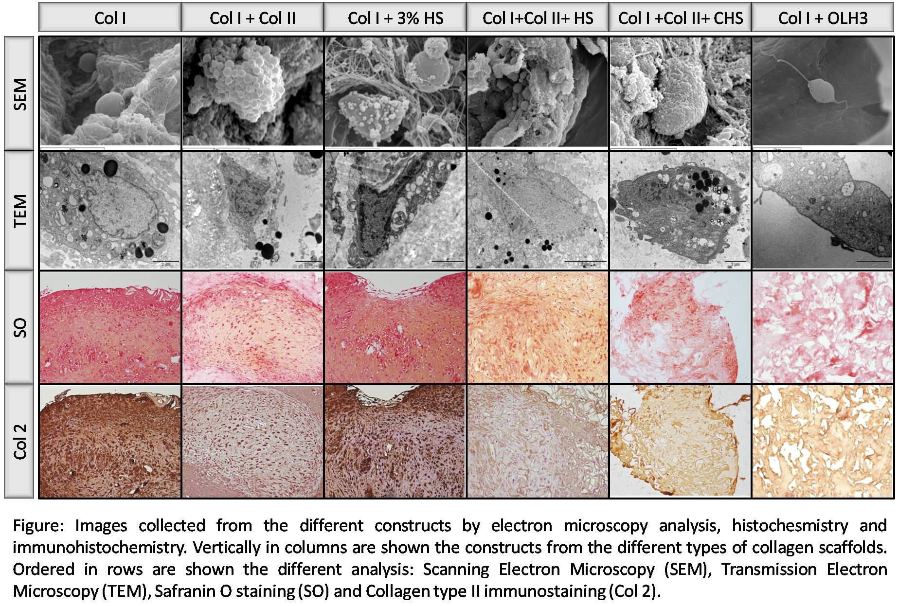
Results: Haematoxilin-Eosin staining showed that hBM-MSCs have grown through the scaffolds. The cellular percentage respect the whole scaffold area was higher than 50% in all the biomaterials and it was observed a big amount of extracellular matrix (ECM) in all of them, except in Col+OLH3. The ECM showed proteoglycans, by means of safranin O (SO) staining, and immunopositivity for Col II in all the constructs, except in Col+OLH3 (Figure). By measurement of relative expression levels (REL) we observed that COLII were expressed by cells in Col I (0.63), Col I+Col II (1.00), Col I+3%HS (0.47), Col I+Col II+HS (0.93), Col I+Col II+CHS (3.57) and Col I+OLH3 (5.20). Moreover, REL of AGG were higher in Col I+Col II+HS (6.47) constructs than in Col I (0.00), Col I+Col II (0.95),Col I+3%HS (0.17), Col I+Col II+CHS (0.00) and Col I+OLH3 (0.00). Electron microscopy showed cells with a big amount of mitochondria and with spherical and oval shapes (Figure). Col released was detected in all supernatant cultures, obtaining the highest concentration (μg/total volume) at all times, in Col I+3%HS cultures except at day 21: Col I (137.33), Col I+Col II+HS (3.33), Col I+Col II (0.00),Col I+3%HS (0.00), Col I+Col II+CHS (13.90) and Col I+OLH3 (0.00) .
Conclusion: Data showed that hBM-MSCs are capable of growing on collagen scaffolds. On HS enriched collagen scaffolds was kept the differentiated phenotype. The ECM characteristics and the gene expression showed the synthesis of a cartilage-like neotissue which could be useful for cartilage tissue engineering. Acknowledgements: Opocrin S.P.A.; CAM (S2009/MAT-1472); CIBER-BBN CB06-01-0040; SAI-UDC; REDICENT; Diputación de A Coruña; Jiménez Díaz Fundation.
Disclosure:
C. Sanjurjo-Rodríguez,
None;
A. H. Martínez-Sánchez,
None;
T. Hermida-Gómez,
None;
I. M. Fuentes-Boquete,
None;
F. J. De Toro,
None;
J. Buján,
None;
S. Díaz-Prado,
None;
F. J. Blanco,
None.
« Back to 2013 ACR/ARHP Annual Meeting
ACR Meeting Abstracts - https://acrabstracts.org/abstract/human-mesenchymal-stem-cells-cultured-on-collagen-scaffolds-for-cartilage-tissue-engineering/

