ACR Meeting Abstracts

ACR Meeting Abstracts

  • Meetings
    • ACR Convergence 2025
    • ACR Convergence 2024
    • ACR Convergence 2023
    • 2023 ACR/ARP PRSYM
    • ACR Convergence 2022
    • ACR Convergence 2021
    • 2020-2009 Meetings
    • Download Abstracts
  • Keyword Index
  • Advanced Search
  • Your Favorites
    • Favorites
    • Login
    • View and print all favorites
    • Clear all your favorites
  • ACR Meetings

Abstract Number: 1846

Integrated spatial and single-cell profiling of treatment-naïve lupus nephritis biopsies

Roopa Madhu1, Rachael Bogle2, DANIEL STEIN3, JIngyi Cao4, Alex Tsoi2, Caleb Marlin5, Thomas Eisenhaure6, Peter Izmirly7, Mala Masson8, Kevin Wei9, Victoria Werth10, Jennifer Barnas11, Michael Brenner12, Judith James5, Johann Gudjonsson13, Joel Guthridge5, Nir Hacohen14, Kelly Ruggles15, Soumya Raychaudhuri16, Jill Buyon17, Michelle Petri18, Brad Rovin19, Andrea Fava20 and ilya Korsunsky16, 1Brigham and Women's Hospital, Brookline, MA, 2University of Michigan, Holland, OH, 3Harvard University, Boston, 4BWH, Boston, 5Oklahoma Medical Research Foundation, Oklahoma City, OK, 6Broad Institute, Cambridge, MA, 7New York University Grossman School of Medicine, New York, NY, 8NYU Langone Health, New York, 9Brigham and Women's Hospital at Harvard Medical School, Boston, MA, 10University of Pennsylvania, Wynnewood, PA, 11University of Rochester, Rochester, NY, 12Brigham and Women's Hospital, Harvard Medical School, Newton, MA, 13University of Michigan, Ann Arbor, MI, 14Broad Institute of MIT Harvard, Cambridge, MA, 15NYU Grossman School of Medicine, Brooklyn, NY, 16Brigham and Women's Hospital, Boston, MA, 17NYU Grossman School of Medicine, New York, NY, 18Johns Hopkins University School of Medicine, Timonium, MD, 19The Ohio State University, Columbus, OH, 20Johns Hopkins University, Baltimore, MD

Meeting: ACR Convergence 2025

Keywords: Lupus nephritis

  • Tweet
  • Click to email a link to a friend (Opens in new window) Email
  • Click to print (Opens in new window) Print
Session Information

Date: Tuesday, October 28, 2025

Title: (1830–1854) Systemic Lupus Erythematosus – Etiology and Pathogenesis Poster

Session Type: Poster Session C

Session Time: 10:30AM-12:30PM

Background/Purpose: Lupus nephritis (LN) is characterized by kidney inflammation and leads to loss of kidney function. Depending on race/ethnicity, up to 60% of Systemic Lupus Erythematosus (SLE) patients develop LN early in their disease. However, the cellular pathways driving disease onset and progression remain poorly defined. Because these pathways involve cell interactions at different spatial scales, spatial mapping of kidney biopsies can provide critical insights. This study aimed to spatially profile kidney biopsies from early LN patients to identify key cellular states, interactions, and regions associated with disease progression and to construct a spatial atlas of early LN pathology.

Methods: We analyzed Formalin-Fixed Paraffin-Embedded (FFPE) kidney biopsies from patients with LN and UPCR between .250 and .499 (n = 8), non-LN SLE (no LN; n = 3), and healthy controls (n = 4). Serial sections were profiled using Xenium (5 µm; 5101 genes) and single-cell FFPE RNA seq (23 µm; whole transcriptome). Xenium transcripts were quality filtered and segmented into cells using a transcript density-based segmentation method (Baysor). Custom pipelines were used to process and integrate Xenium and scFFPE data. After QC, we obtained ~400,000 cells from Xenium and ~100,000 cells from scFFPE.

Results: Integration of scFFPE and Xenium data revealed 8 major cell lineages based on marker expression (Fig. 1A, B). LN and non-LN samples showed enrichment of lymphocytes compared to controls (Fig. 1C). Cell projections (Xenium) confirmed that profiled cells localized to expected anatomical regions (Fig. 1D). Using the public AMP SLE reference and in house pipelines, we annotated 43 cell states across the 8 cell lineages and mapped in tissue space. Transcript-based tissue segmentation with Tessera identified six regions: 3 tubular, 1 glomerular, and two interstitial (Fig. 2A, B).We next quantified cell state enrichment in each region across groups (Fig. 3). In controls, cell states localized to expected regions. In LN, macrophage subsets M5 (GPNMB high NUPR high) and M7 (SPP1 high FABP5 high) were enriched in glomerular regions, compared to interstitial enrichment in controls and non-LN SLE. Colocalization analyses revealed that M5 and M7 colocalized in the glomerular regions in LN samples, suggesting recruitment to inflamed regions during early disease.

Conclusion: We generated a comprehensive kidney spatial atlas of early LN, non-LN SLE, and healthy controls, integrating scFFPE RNA sequencing and Xenium spatial transcriptomics. In this initial study of patients with early-stage LN, we have started to characterize distinct cell states that may be involved in the development of LN in patients with SLE. We found that patients with SLE and no LN accumulated lymphocytes, suggesting that even in the absence of frank LN, the kidneys of lupus patients may be affected. We also found that resident macrophages tend to move from the interstitium to the glomerular region in patients with early LN. These macrophages may be involved in initiating glomerular injury in LN.
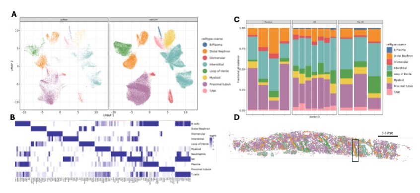
Supporting image 1Figure1: (A) UMAP projection of cells from scFFPE RNA sequencing and Xenium datasets. Cells are colored by their major cell types. (B) Heat map of logFC of top 15 marker genes for every cell type. (C) Relative abundance of cell type populations in each sample. Relative abundance is the ratio of number of cells of each type to the total number of cells in the sample. (D) Spatial projection of the cell types in (A) onto tissue space. Insert highlights the distal tubule.

Supporting image 2Figure2: (A) Spatial segmentation of the tissue shown in 1D. Regions are colored by their type. Insert highlights the distal tubule in 1D, which is now detected and annotated as a DT rich region. (B) Relative abundance of cell types in each region in each sample in the dataset. Regions are annotated based on their cell composition.

Supporting image 3Figure3: (A-C): Log enrichment of cell states within each spatial region: (A) in control samples, (B) No LN samples, (C) LN samples.


Disclosures: R. Madhu: None; R. Bogle: None; D. STEIN: None; J. Cao: None; A. Tsoi: Galderma, 5, Johnson and Johnson, 5; C. Marlin: None; T. Eisenhaure: None; P. Izmirly: Hansoh Bio, 2; M. Masson: None; K. Wei: 10X Genomics, 5, anaptysbio, 2, Gilead, 5, Merck/MSD, 5, Mestag, 2, Pfizer, 2; V. Werth: AbbVie/Abbott, 2, Amgen, 2, 5, Anaptysbio, 2, Argenx, 2, 5, AstraZeneca, 2, 5, Biogen, 2, 5, Boehringer-Ingelheim, 2, Bristol-Myers Squibb(BMS), 2, Cabaletta Bio, 2, Calyx, 2, CSL Behring, 5, Cugene, 2, Eli Lilly, 2, EMD Serona, 2, 2, Evommune, 2, Gilead, 2, 5, GlaxoSmithKlein(GSK), 2, Horizon, 5, Immunovant, 2, Janssen, 2, Kwoya Kirin, 2, Merck/MSD, 2, Novartis, 2, Nuvig, 2, Pfizer, 2, 5, Regeneron, 2, 5, Roche, 2, Rome Pharmaceuticals, 5, Sanofi, 2, Takeda, 2, UCB, 2, 2, Ventus, 2, Viela, 5; J. Barnas: None; M. Brenner: AbbVie/Abbott, 2, Johnson & Johnson, 5, Mestag Therapeutics, 2, 8, Moderna, 2, Ono Pharma, 5, Simcere, 2; J. James: GlaxoSmithKlein(GSK), 2, Progentec, 5; J. Gudjonsson: None; J. Guthridge: None; N. Hacohen: Calico Biosciences, 5, Cytoreason, 1, 11, Danger Bio, 1, 11, Moderna, 5, Repertoire Immune Medicines, 1, 11, Takeda, 5; K. Ruggles: None; S. Raychaudhuri: Bonito, 1, Bristol-Myers Squibb(BMS), 1, Janssen, 1, Mestag, 1, 8, Nimbus, 2, Pfizer, 1, Sonoma, 1, 8, Visterra, 6; J. Buyon: Artiva Biotherapeutics, 2, Biogen, 2, Bristol-Myers Squibb(BMS), 2, Celgene, 2, CLIMB Bio Operating, 2, GlaxoSmithKlein(GSK), 2, Janssen, 2, Related Sciences, 2, UCB, 2; M. Petri: Amgen, 2, AnaptysBio, 2, Annexon Bio, 2, AstraZeneca, 2, 5, Atara Biosciences, 2, Aurinia, 2, 5, Autolus, 2, Bain Capital, 2, Baobab Therapeutics, 2, Biocryst, 2, Biogen, 2, Boxer Capital, 2, Cabaletto Bio, 2, Caribou Biosciences, 2, CTI Clinical Trial and Consulting Services, 2, CVS Health, 2, Dualitybio, 2, Eli Lilly, 2, 5, EMD Serono, 2, Emergent, 2, Escient Pharmaceuticals, 2, Exagen, 5, Exo Therapeutics, 2, Gentibio, 2, GlaxoSmithKlein(GSK), 2, 5, iCell Gene Therapeutics, 2, Innovaderm Research, 2, IQVIA, 2, Janssen, 5, Kezar Life Sciences, 2, Kira Pharmaceuticals, 2, Nexstone Immunology, 2, Nimbus Lakshmi, 2, Novartis, 2, Ono Pharma, 2, PPD Development, 2, Proviant, 2, Regeneron, 2, Seismic Therapeutic, 2, Senti Biosciences, 2, Sinomab Biosciences, 2, Steritas, 2, Takeda, 2, Tenet Medicines, 2, TG Therapeutics, 2, UCB, 2, Variant Bio, 2, Worldwide Clinical Trials, 2, Zydus, 2; B. Rovin: Alexion, 2, Artiva, 2, 11, AstraZeneca, 2, Aurinia, 2, 5, Biogen, 2, 5, Bristol Myers Squibb, 2, Cabelleta, 2, Century, 2, F. Hoffman-La Roche Ltd/Genentech, Inc., 2, GlaxoSmithKlein(GSK), 2, Novartis, 2; A. Fava: Artiva, 2, AstraZeneca, 1, 2, Bain Capital, 2, Biogen, 1, Bristol-Myers Squibb(BMS), 2, Exagen, 5, 9, Quotient Therapeutics, 2, UCB, 6, Zenas, 2; i. Korsunsky: Mestag therapeutics, 2.

To cite this abstract in AMA style:

Madhu R, Bogle R, STEIN D, Cao J, Tsoi A, Marlin C, Eisenhaure T, Izmirly P, Masson M, Wei K, Werth V, Barnas J, Brenner M, James J, Gudjonsson J, Guthridge J, Hacohen N, Ruggles K, Raychaudhuri S, Buyon J, Petri M, Rovin B, Fava A, Korsunsky i. Integrated spatial and single-cell profiling of treatment-naïve lupus nephritis biopsies [abstract]. Arthritis Rheumatol. 2025; 77 (suppl 9). https://acrabstracts.org/abstract/integrated-spatial-and-single-cell-profiling-of-treatment-naive-lupus-nephritis-biopsies/. Accessed .
  • Tweet
  • Click to email a link to a friend (Opens in new window) Email
  • Click to print (Opens in new window) Print

« Back to ACR Convergence 2025

ACR Meeting Abstracts - https://acrabstracts.org/abstract/integrated-spatial-and-single-cell-profiling-of-treatment-naive-lupus-nephritis-biopsies/

Advanced Search

Your Favorites

You can save and print a list of your favorite abstracts during your browser session by clicking the “Favorite” button at the bottom of any abstract. View your favorites »

Embargo Policy

All abstracts accepted to ACR Convergence are under media embargo once the ACR has notified presenters of their abstract’s acceptance. They may be presented at other meetings or published as manuscripts after this time but should not be discussed in non-scholarly venues or outlets. The following embargo policies are strictly enforced by the ACR.

Accepted abstracts are made available to the public online in advance of the meeting and are published in a special online supplement of our scientific journal, Arthritis & Rheumatology. Information contained in those abstracts may not be released until the abstracts appear online. In an exception to the media embargo, academic institutions, private organizations, and companies with products whose value may be influenced by information contained in an abstract may issue a press release to coincide with the availability of an ACR abstract on the ACR website. However, the ACR continues to require that information that goes beyond that contained in the abstract (e.g., discussion of the abstract done as part of editorial news coverage) is under media embargo until 10:00 AM CT on October 25. Journalists with access to embargoed information cannot release articles or editorial news coverage before this time. Editorial news coverage is considered original articles/videos developed by employed journalists to report facts, commentary, and subject matter expert quotes in a narrative form using a variety of sources (e.g., research, announcements, press releases, events, etc.).

Violation of this policy may result in the abstract being withdrawn from the meeting and other measures deemed appropriate. Authors are responsible for notifying colleagues, institutions, communications firms, and all other stakeholders related to the development or promotion of the abstract about this policy. If you have questions about the ACR abstract embargo policy, please contact ACR abstracts staff at [email protected].

Wiley

  • Online Journal
  • Privacy Policy
  • Permissions Policies
  • Cookie Preferences

© Copyright 2025 American College of Rheumatology