Session Information
Date: Tuesday, October 28, 2025
Title: Abstracts: Spondyloarthritis Including Psoriatic Arthritis – Basic Science (1752–1757)
Session Type: Abstract Session
Session Time: 10:15AM-10:30AM
Background/Purpose: Psoriatic arthritis (PsA) is a complex and heterogeneous inflammatory disorder involving the axial skeleton in approximately 30% of patients. The mechanisms that underlie psoriatic axial inflammation (AxPsA) are not well understood and longitudinal studies to examine the immune mechanisms involved in disease pathogenesis and progression using human cells in an animal model have not been performed. In previous analyses, we demonstrated that human PsA phenotypes in the peripheral skeleton, entheses and skin are recapitulated in the NSG-GM3 humanized mouse model. Herein, we examined if axial disease is phenocopied in this mouse model.
Methods: Two groups of NSG-SGM3 mice were injected with serum and peripheral blood mononuclear cells (PBMC) collected from three biologic naive PsA patients with inflammatory back pain and radiographic or MRI documented sacroiliitis (AxPsA) and controls (PsA patients without axial disease). At day 30, mice were anesthetized, and their spines were dissected for radiographic and micro-CT scan analysis. Tissue sections from decalcified spines were stained with anti CD45, CD14, CD8, Ki67, TNF and Il-17 antibodies.
Results: NSG-SGM3 mice injected with serum and engrafted with PBMC from patients with AxPsA exhibited pronounced spinal deformities. Radiographic imaging revealed marked cervical and thoracic kyphosis, bone erosion and osteoproliferation in the tail and micro-computed tomography (micro-CT) scans identified both bone resorption and pathological bone fusion in the sacroiliac joint and lumbar spine. In contrast, mice injected with PBMC and sera from non-AxPsA controls did not demonstrate significant inflammation or structural alterations. Histological analysis in the spine of mice injected with AxSpA PBMC and sera revealed accumulation of lymphocytes in and around the dorsal root ganglion (DRG) and in the surrounding muscle. Inflammatory cell infiltrates were characterized by proliferating CD3 T and CD8 T cells that expressed both TNF and IL-17. TNF and IL-17 expression were elevated most notably in the DRG.
Conclusion: Injection of PBMC and sera from AxPsA patients recapitulated axial clinical features in hu PsA mice. HuAxPsA mice developed kyphosis in the distal cervical and thoracic spine and structural damage in the lower lumbar and sacroiliac region and tail, including bone loss and new bone formation. Proliferating CD3+ T and CD8+ T cells expressing TNF and IL-17 were noted near the DRG and in surrounding tissues and paravertebral muscles. This model will accelerate investigation of immune mechanisms in AxPsA.
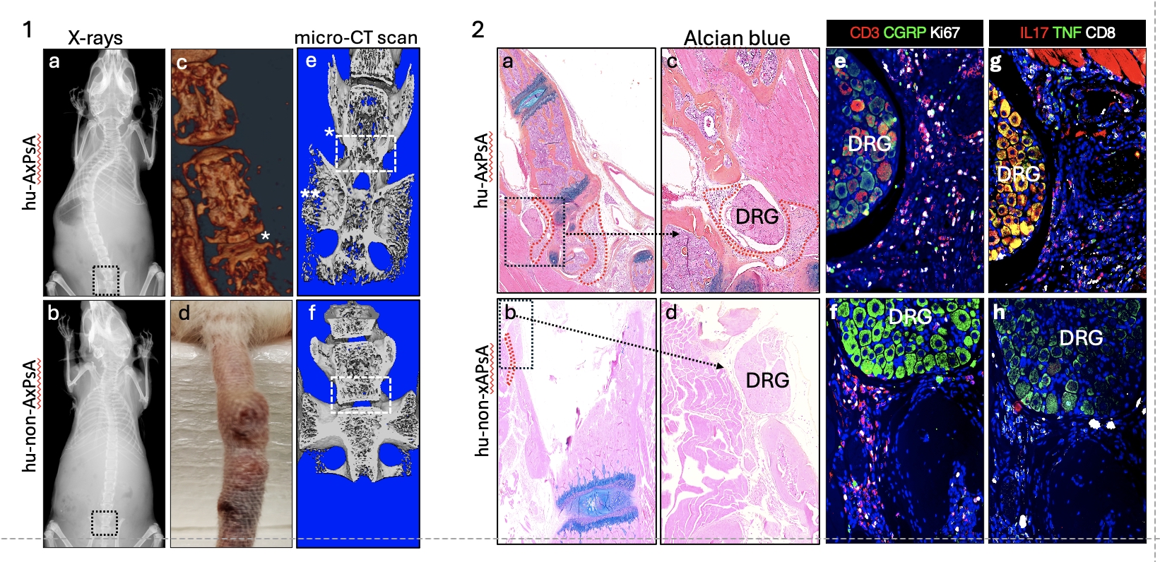 Figure 1. Humanized NSG-SGM3 mice generated with sera and PBMC from AxPsA and non-AxPA patients. 1) Hu-AxPsA mice demonstrated (a), cervical and thoracic kyphosis not seen in controls (b) (c), spinal fusion and erosion in the lumbar spine (c, * fusion and erosion below), (d) tail inflammation and necrosis and (e) L5, S1 fusion * and bone resorption ** not observed in controls (f). Figure 2. Alcian blue staining of the spine shows abundant infiltrating immune cells (dotted red line) around the dorsal root ganglion (DRG) (a,c) and low infiltration in the spine of hu-non-AxPsA mice (b,d). Immunofluorescence staining shows more proliferating CD3 (red and white) and IL-17+ (red) TNF + (green) CD8+ (white) and Il-17+ TNF+ cells in the DRG of hu-AxPsA (e, g) compared to hu-non-AxpSA mice (f,h).
To cite this abstract in AMA style:
Ritchlin C, Rangel-Moreno J, Rangel-Garcia A, Krantz M, Brien M, Sathilaseenlan T, Fox J, Wood R, Garcia-Hernandez M. Axial Psoriatic Arthritis is Phenocopied in a Humanized Mouse Model [abstract]. Arthritis Rheumatol. 2025; 77 (suppl 9). https://acrabstracts.org/abstract/axial-psoriatic-arthritis-is-phenocopied-in-a-humanized-mouse-model/. Accessed .« Back to ACR Convergence 2025
ACR Meeting Abstracts - https://acrabstracts.org/abstract/axial-psoriatic-arthritis-is-phenocopied-in-a-humanized-mouse-model/