Session Information
Session Type: Abstract Submissions (ACR)
Background/Purpose: Systemic lupus erythematosus (SLE) is a multisystem disease, which affects mostly women at reproductive age, and can promote premature ovarian dysfunction related to factors associated to rheumatic disease or its treatment. The assessment of indicators of ovarian reserve can determine if there are differences between these patients and the general population, prior to the establishment of the climacteric. The aim of this study was to evaluate if there are differences in ovarian reserve markers in systemic lupus erythematosus (SLE) patients compared to controls, and explore the relationship of such markers with clinical and treatment features of SLE patients, in addition to inflamatory activity (SLEDAI/ACR) and damage (SLICC/ACR-DI) disease scores.
Methods: This was a controlled cross-sectional study including 27 women with SLE and 27 controls. All participants were between 18 and 40 years, were eumenorrheic and did not use hormone therapy or hormone contraceptives in the past 6 months. Clinical data were assessed at a regular follow up visit, serum concentrations of follicle stimulating hormone (FSH) and anti-mullerian hormone (AMH), and through transvaginal ultrasound antral follicle count were assessed at early follicular phase of a subsequent menstrual cycle.
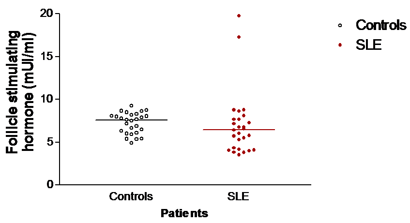
Results: Mean age of SLE patients was 30.9 years (SD±4.8) and had 102.7 months (SD±66.7) of lenth disease. We found no difference between SLE group and control group at analysis of AFC [median (interquartile interval) 7 (5 – 13) vs. 11 (7 – 12), p=0.076], FSH [6.44 (4.19 – 7.69) vs. 7.5 (6.03 – 8.09) mUI/ml, p=0.135] and AMH levels [1.23 (0.24 – 4.63) ng/ml vs. 1.52 (1.33 – 1.88) ng/ml, p=0.684]. However, AMH values in SLE group were more heterogeneous compared to control group. The presence of nephritis and the cumulative dose of cyclophosphamide were factors individually related to reduced ovarian reserve, by association with lower values of AFC and AMH. At multivariate logistic regression, control group was more likely to have higher AMH values than the SLE (OR 5.2, 95% CI 1.286 – 20.405, p=0.021) and in the SLE group, AMH was associated with lower maximum corticosteroid doses in the follow-up (OR 0.95, 95%CI 0.894-1.000, p=0.50). AFC was associated with lower scores of SLICC/ACR-DI (OR: 0.14, 95% CI 0.025-0.841, p=0.031).
Conclusion: SLE patients who were eumenorrheic had average values of ovarian reserve markers similar to controls. However, AMH had a wide range of values in that group, requiring study of other markers to clarify the best clinical application for it. Ovarian function is more compromised in patients with nephritis, high cumulated dose of cyclophsphamide and higher disease damage scores.
Figure . Scatters showing comparison of ovarian reserve markers in SLE patients between and controls in medians: AFC (Figure A), FSH (Figure B), and AMH (Figure C).
Disclosure:
O. B. Malheiro,
None;
C. P. Rezende,
None;
G. A. Ferreira,
None;
F. M. Reis,
None.
« Back to 2012 ACR/ARHP Annual Meeting
ACR Meeting Abstracts - https://acrabstracts.org/abstract/ovarian-reserve-markers-in-reproductive-age-women-with-systemic-lupus-erythematosus/


