Session Information
Session Type: Abstract Submissions (ACR)
Background/Purpose: Neurocognitive dysfunction (NCD) is common in childhood-onset systemic lupus erythematosus (cSLE), and often difficult to detect with current resources. Diffusion tensor imaging (DTI) is a magnetic resonance imaging technique, which allows the calculation of various measures of tissue microstructural integrity, and structural connectivity via evaluation of white matter tracts. The objectives were to use DTI to investigate specific anatomic changes in the brain in cSLE patients with and without NCD in comparison to controls, and to assess the potential of these measures for use as imaging biomarkers.
Methods: Formal neuropsychological testing using the cSLE Neurocognitive Battery (Ross et al, 2010) was done to measure cognitive ability and identify NCD in cSLE. DTI was performed in cSLE patients with NCD (cSLE-NCD; n = 6), without NCD (cSLE-noNCD; n = 9) and healthy controls (n = 14). DTI was acquired on a 3 Tesla scanner at 2mm isotropic resolution for 32 gradient directions using an echo-planar imaging protocol with TR/TE = 8800/88 ms and a b-factor of 1000 s/mm2. DTI data were processed using FSL 4.1 (http://www.fmrib.ox.ac.uk/fs/) including eddy current correction. Diffusion tensors were calculated using the Diffusion Toolkit (http://trackvis.org/blog/tag/diffusion-toolkit/), determining the principal diffusion direction per voxel throughout the white matter. This resulted in a 3D map of fiber streamlines (FS). Structural connectivity between two brain regions was defined as the number of FS with an endpoint in each of those regions. Fiber density was defined as the number of FS passing through a region regardless of endpoints. The entire brain was divided into 116 distinct regions using a standard anatomical atlas (Tzourio-Mazoyer et al, 2002). A binary matrix summarized structural network architecture by setting a connectivity threshold at > 20 FS. Group comparisons were made for density and probability of connection to each region individually and across the entire brain.
Results: A significant decrease in density was observed for the cSLE-NCD group vs. controls (p = 0.002) or cSLE-noNCD (p= 0.001). No difference in density was found between controls and cSLE-noNCD. Connectivity over the entire brain and individual regions followed a similar pattern of significant decrease for the cSLE-NCD group.
Conclusion: Based on DTI, neurocognitive deficits in cSLE are associated with loss of fiber density and interregional connectivity that suggest breakdown of structural network integration. These results complement our previously reported functional and volumetric findings (DiFrancesco et al, 2013; Gitelman et al, 2013), suggesting that cSLE-NCD is associated with measurable changes in grey and white matter. If confirmed in larger cohorts, DTI abnormalities may be used as imaging biomarkers of cSLE-associated NCD.
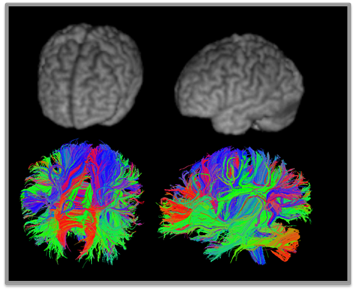
Figure 1: Tractography in single subject
Disclosure:
J. T. Jones,
None;
M. DiFrancesco,
None;
A. I. Zaal,
None;
M. S. Klein-Gitelman,
None;
D. Gitelman,
None;
J. Ying,
None;
H. Brunner,
No,
2.
« Back to 2013 ACR/ARHP Annual Meeting
ACR Meeting Abstracts - https://acrabstracts.org/abstract/childhood-onset-systemic-lupus-erythematosus-with-clinical-neurocognitive-dysfunction-have-lower-nodal-density-and-connectivity-on-diffusion-tensor-imaging/

