Session Information
Date: Monday, November 18, 2024
Title: Systemic Sclerosis & Related Disorders – Basic Science Poster II
Session Type: Poster Session C
Session Time: 10:30AM-12:30PM
Background/Purpose: Our previous single-cell RNA-sequencing study identified two major fibroblast subpopulations in healthy skin biopsies: SFRP2/DPP4 and FMO1/LSP1 fibroblasts¹. Additionally, we discovered that adventitial fibroblasts are comprised of two primary clusters based on APOE expression levels: APOE^hi/CCL19/C7-expressing fibroblasts and APOE^low/FMO1/MYOC-expressing fibroblasts. We showed that FMO1/MYOC-expressing fibroblasts exhibit both perivascular and interstitial distribution, whereas CCL19-secreting fibroblasts are predominantly perivascular2. Although different studies suggest the importance of vascular injury in the pathophysiology of scleroderma (SSc), modifications in adventitial fibroblasts and their morphological changes remain poorly understood. Here, we aim to study the changes in the distribution patterns of adventitial fibroblasts in healthy skin samples compared to SSc skin biopsies.
Methods: Forearm skin biopsies were obtained from 12 healthy individuals and 8 scleroderma patients following an approved protocol by the Institutional Review Board of the University of Pittsburgh School of Medicine. Paraffin-embedded skin samples were used for double and triple immunofluorescence staining, using Tyramide amplification and various antibodies targeting adventitial fibroblast markers such as CCL19, MYOC, and FMO1. Confocal microscopy was performed using Olympus FluoView 3000.
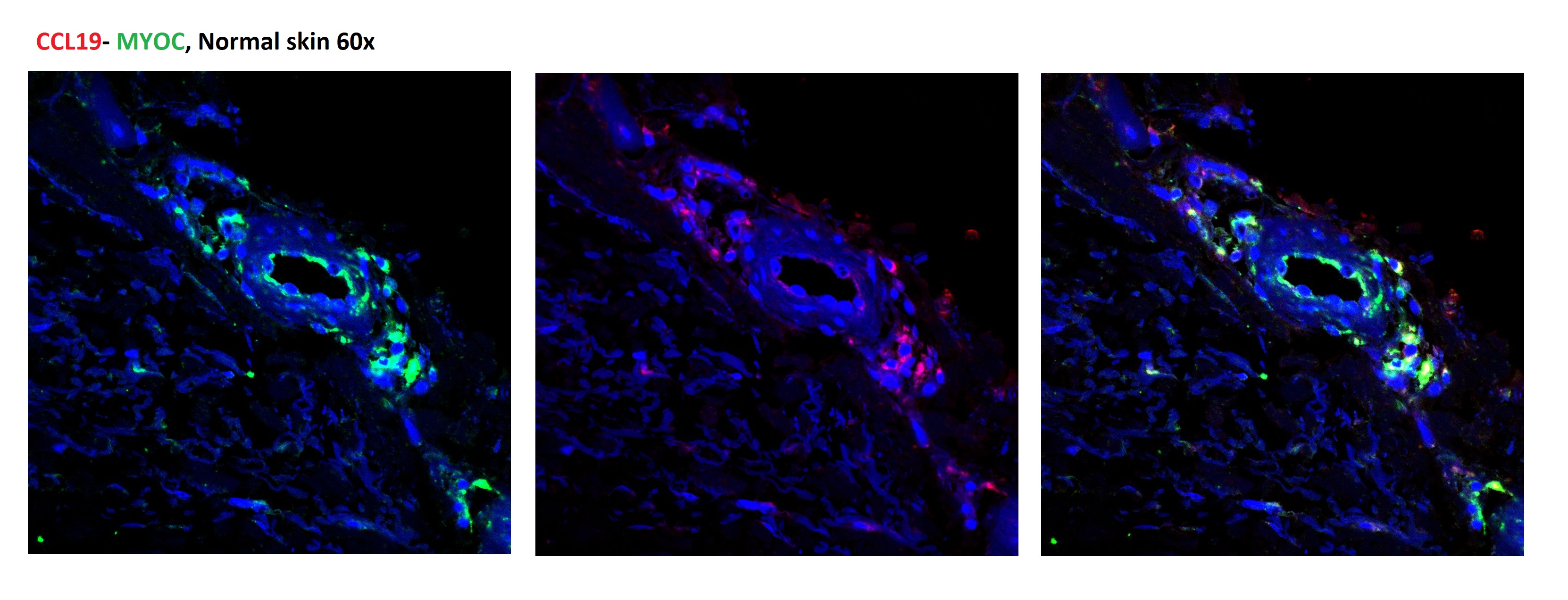
Results: Our analysis revealed dramatic changes in the distribution of adventitial fibroblasts between healthy and SSc skin samples. In healthy skin, CCL19 and MYOC stained overlapping populations of adventitial fibroblasts. In SSc skin, we observed a marked increase in CCL19-expressing fibroblasts, predominantly around the vasculature with extensions into the interstitium adjacent to vascular structures. These cells appeared to be denser in the upper dermis, particularly the papillary dermis. In patients with advanced SSc, this expansion into interstitium was more pronounced, while in healthy individuals the distribution pattern of CCL19 fibroblasts remained limited to the perivascular regions. Similarly, FMO1 and MYOC-expressing fibroblasts exhibited expanded interstitial presence in SSc skin, following the pattern observed in CCL19 expansion. Interestingly, MYOC-positive cells also showed increased presence in the papillary dermis of SSc skin, mirroring the distribution of CCL19 fibroblasts.
Conclusion: Our study indicates complex patterns of adventitial fibroblasts, expressing overlapping markers. These fibroblast populations appear to infiltrate the interstitium in the skin of SSc patients compared to healthy samples. These differences along with the increase in specific fibroblast subpopulations suggest that changes in adventitial fibroblasts may have a critical role in modifying vascular integrity and perivascular inflammation. These studies using protein markers confirm and extend our previous observations, characterizing various adventitial subpopulations by single-cell RNA sequencing. Future studies using spatial transcriptomics will provide further insights into the role of changes in different fibroblast subpopulations in SSc skin.
To cite this abstract in AMA style:
Nazari B, Tabib T, Morse C, Lafyatis R. Distribution and Morphological Changes of Adventitial Fibroblasts in Healthy and Scleroderma Skin [abstract]. Arthritis Rheumatol. 2024; 76 (suppl 9). https://acrabstracts.org/abstract/distribution-and-morphological-changes-of-adventitial-fibroblasts-in-healthy-and-scleroderma-skin/. Accessed .« Back to ACR Convergence 2024
ACR Meeting Abstracts - https://acrabstracts.org/abstract/distribution-and-morphological-changes-of-adventitial-fibroblasts-in-healthy-and-scleroderma-skin/


