Session Information
Date: Saturday, November 16, 2024
Title: Metabolic & Crystal Arthropathies – Basic & Clinical Science Poster I
Session Type: Poster Session A
Session Time: 10:30AM-12:30PM
Background/Purpose: Calcium pyrophosphate deposition disease is a common condition that can cause chronic polyarticular inflammatory joint disease, which sometimes resembles rheumatoid arthritis. Misdiagnosis is especially common in the case of seronegative rheumatoid arthritis and can lead to unnecessary treatment with potentially toxic medications. Prior investigations aiming to estimate the prevalence of CPPD in patients with seronegative rheumatoid arthritis utilized conventional radiography. Our goal was to determine the proportion of patients initially suspected to have or diagnosed with seronegative rheumatoid arthritis that could meet classification criteria for calcium pyrophosphate crystal deposition with the aid of ultrasonography.
Methods: The investigation involved a retrospective chart review of the patients who visited our dedicated rheumatology musculoskeletal ultrasound (MSKUS) clinic at the Birmingham Veterans Affairs Medical Center (BVAMC) between 01/01/2020 and 12/31/2023. The cases of a suspected or possible diagnosis of seronegative rheumatoid arthritis were identified. The results of MSKUS were reviewed for the presence of calcium pyrophosphate deposition within fibrocartilage and hyaline cartilage according to the OMERACT definitions. For each one of the patients we also calculated the total score according to the 2023 ACR/EULAR classification criteria for CPPD disease.
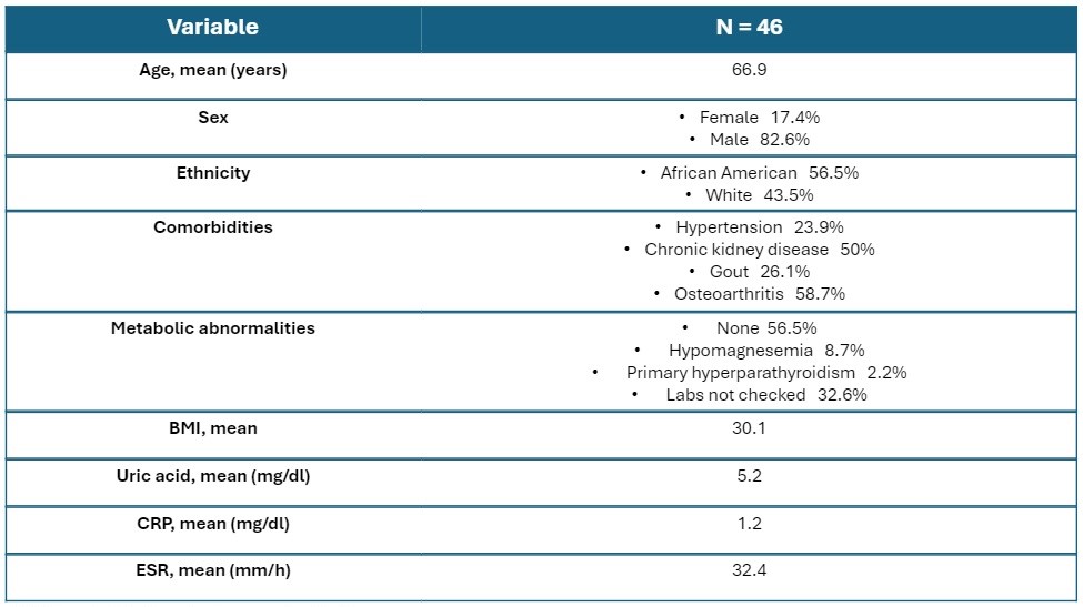
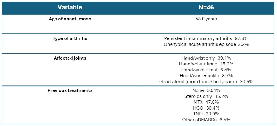
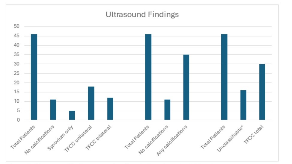
Results: Among 316 patients who visited the BVAMC musculoskeletal ultrasound clinic between 01/01/2020 and 12/31/2023, 46 were found to have a possible or established diagnosis of seronegative rheumatoid arthritis prior to ultrasonography. The majority of patients were male (82.6%), and many of them had previously been treated with disease-modifying agents. Baseline characteristics are shown in Tables 1 and 2. Most of the patients (95.7%) had MSKUS performed on hands and wrists. 35/46 (76.1%) were found to have some degree of calcifications on MSKUS, with 30/46 (65.2%) showing calcium deposition within the triangular fibrocartilage complex of the wrist (TFCC – a classic location for CPPD disease) and 5/46 (10.9%) – within the synovium of the joints only. 13/46 (28.2%) patients were classified as having CPPD disease according to the 2023 ACR/EULAR scoring system. For 22/46 (47.8%) patients fibrocartilage calcifications were detected by ultrasonography but not with conventional radiographs. The main results are shown in Figure 1.
Conclusion: A significant proportion of patients with suspected diagnosis of seronegative rheumatoid arthritis had evidence of calcium pyrophosphate deposition in the joints as detected by ultrasonography. Approximately one-quarter of cases could be reclassified as CPPD by validated criteria, leading to a possible reconsideration of the diagnosis. We believe the results of our study might improve the understanding of this common crystalline arthropathy and encourage rheumatologists to utilize musculoskeletal ultrasound as a diagnostic tool in challenging cases to help guide treatment decisions and limit therapies with potential side effects when not clinically indicated.
*Unclassifiable – no calcifications or synovium only (not in the ACR/EULAR classification criteria)
To cite this abstract in AMA style:
Potera J, Gaffo A, Day A, Urquiaga M, Alexander A. Identification of Calcium Pyrophosphate Deposition Using Ultrasonography in Patients with a Suspected Diagnosis of Seronegative Rheumatoid Arthritis [abstract]. Arthritis Rheumatol. 2024; 76 (suppl 9). https://acrabstracts.org/abstract/identification-of-calcium-pyrophosphate-deposition-using-ultrasonography-in-patients-with-a-suspected-diagnosis-of-seronegative-rheumatoid-arthritis/. Accessed .« Back to ACR Convergence 2024
ACR Meeting Abstracts - https://acrabstracts.org/abstract/identification-of-calcium-pyrophosphate-deposition-using-ultrasonography-in-patients-with-a-suspected-diagnosis-of-seronegative-rheumatoid-arthritis/


