Session Information
Date: Monday, November 13, 2023
Title: (1221–1255) Pediatric Rheumatology – Clinical Poster II: Connective Tissue Disease
Session Type: Poster Session B
Session Time: 9:00AM-11:00AM
Background/Purpose: There is a critical need for tools to distinguish between maturational changes mimicking subchondral edema from pathologic inflammation within the SI joints on MRI. We aimed to establish standards for interpretation of diffusion weighted imaging (DWI) in healthy children.
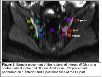
Methods: This was a retrospective cross-sectional study of MRI scans of healthy controls identified from a prior study that evaluated the appearance of MRI in healthy children (Chauvin et. al, 2019) and through query of the digital archiving system at a single center across a range of bone maturation. DWI and short-tau inversion recovery (STIR) images were analyzed independently by 2 pediatric radiologists using custom software (Parametric MRI, Philadelphia, PA; https://www.parametricmri.com/). DWI series performed in the axial plane were reformatted by the software into a semi-coronal plane. To establish normative ranges of apparent diffusion coefficient (ADC) in controls, circular regions of interest (ROIs) of 0.15-0.22 cm2 were placed at the superior, mid, and inferior portions of the anterior, mid, and posterior ilium and sacrum along the cartilaginous SI joint (Figure 1), avoiding sclerosis. ADC was calculated using a linear log model and all available b values. The type (I-IV) of sacral apophyseal/periarticular maturational signal was recorded. Mixed effects regression models with subject-specific random effect to account for clustering were used to determine if the ROIs (average ADC from the 2 raters) for the superior, mid, and inferior locations for the anterior, mid, posterior portions of the sacrum and ilium differed significantly for each maturational type.
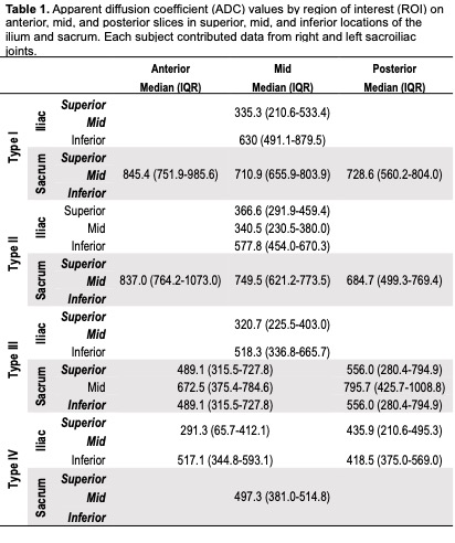
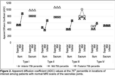
Results: DWI and STIR sequences from 63 healthy children were assessed. Table 1 lists the ADC for each part of the SI joint for maturational types I-IV. Figure 2 shows the 75th percentile of normative values. ADC at the superior, mid, and inferior portions of the anterior, mid, and posterior of the sacrum were not significantly different for type I, II, and IV maturation; for type III, ADC from the posterior sacrum was significantly higher than the anterior and mid slices. The median ADC values in the sacrum were significantly different between maturational types (p < 0.001) and decreased from type I to type IV. ADC values were significantly different in the ilium between the inferior, mid, and superior parts of the SI joint (p < 0.001) with higher ADC values observed in the inferior locations compared to the superior or mid portion of the joint in anterior, mid, and posterior slices for all 4 maturational types.
Conclusion: We established ADC normative values for the superior, mid, and inferior portions of the anterior, mid, and posterior sacrum and ilium along the cartilaginous SI joint by maturational type. Observed trends with more variable ADC in the ilium and higher ADC at the inferior aspect of the ilium are in accordance with prior observations on STIR in the maturing SI joint; however, the ADC from DWI goes beyond prior STIR observations by quantifying the difference(s). Next steps are to establish empirical cut-offs to distinguish normal maturational changes from pathologic inflammation to improve the specificity and confidence in the diagnostic interpretation of the MRI.
To cite this abstract in AMA style:
Francavilla M, Khrichenko D, Brandon T, Chauvin N, Xiao R, Weiss P. Apparent Diffusion Coefficient (ADC) from Diffusion Weighted Imaging (DWI) as an Objective Tool to Differentiate Normal Maturational Subchondral Signal from Inflammation at the Sacroiliac Joint in Youth [abstract]. Arthritis Rheumatol. 2023; 75 (suppl 9). https://acrabstracts.org/abstract/apparent-diffusion-coefficient-adc-from-diffusion-weighted-imaging-dwi-as-an-objective-tool-to-differentiate-normal-maturational-subchondral-signal-from-inflammation-at-the-sacroiliac-joint-in-you/. Accessed .« Back to ACR Convergence 2023
ACR Meeting Abstracts - https://acrabstracts.org/abstract/apparent-diffusion-coefficient-adc-from-diffusion-weighted-imaging-dwi-as-an-objective-tool-to-differentiate-normal-maturational-subchondral-signal-from-inflammation-at-the-sacroiliac-joint-in-you/


