Session Information
Session Type: Abstract Session
Session Time: 2:00PM-3:30PM
Background/Purpose: Ultrasonography has been validated as a diagnostic tool for giant cell arteritis (GCA). There is a need to develop training resources, because the glucocorticoid responsiveness of abnormalities of affected vessels and the uunpredictability and relative scarcity of presentation makes it challenging to predictably use real patients for educational purposes. To address this, we have developed cost-effective ultrasound phantoms using high-resolution 3D printing techniques [1]. The aim of this study is to validate these models within the subgroup on large vessel vasculitis of the Outcome Measures in Rheumatology (OMERACT) ultrasound working group.
Methods: Normal and pathological phantoms of the axillary (AA) and temporal artery (TA) were developed and printed using a Formlabs SLA 3D printer. The phantoms were then embedded in a special gelatine as eight sets of three (AA) or two (TA). Participants from twelve European countries were provided with eight sets of both AA and TA phantoms. The phantoms were evaluated individually in a blinded fashion using a predefined protocol and the OMERACT definitions of abnormalities in GCA [2], [3]. The artificial intima-media thickness (IMT) of each vessel phantom was measured at most prominent site in mm, preferably on the distal wall [4]. The phantoms were classified by each investigators normal (AA&TA), acute (AA&TA), chronic (AA only), or none of the above (AA&TA). Descriptive parameters and interrater-reliability were calculated. Furthermore, we conducted a one-way analysis of variance.
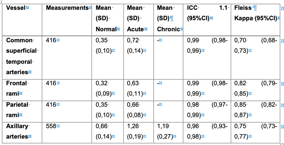
Results: The phantoms were correctly classified in 87% of cases resulting in a Fleiss’ kappa of 0.80 (95%CI 0.78-0.82). The published cut-off values for normal and pathological IMT were met, with the exception of the parietal branch of the TA [5]. Excellent agreement among participants was demonstrated for IMT measurements, with an intraclass correlation coefficient (ICC 1.1) of 0.98 (95% CI 0.98-0.99). The results of IMT measurements are shown in Table 1. A one-way analysis of variance (ANOVA) revealed a significant difference in average IMT between pathological and healthy phantoms for both AA and TA. Detailed results of the ANOVA, divided by the different arteries, are presented in Table 2.
Conclusion: Our novel ultrasound models accurately represent the normal state and pathology of GCA adhering to the OMERACT definitions and cut-off values for IMT. There was a high agreement among investigators to correctly classify the models and good reliability with measurement of artificial IMT. This is also a first step in the evaluation of the phantom for educational purposes.
Notice, that there are no chronic phantoms of temporal arteries (SD: standard deviation; ICC: intra-class correlation coefficient; CI: confidence interval)
The table shows the results of the analysis of variance along with their significance (p-value) and their effect size (η2) (p-value: probability value; η2: partial eta square; df: degrees of freedom; CI: confidence interval)
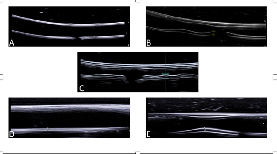
A=Normal axillary artery; B=Axillary artery with acute GCA pathology; C=Axillary artery with GCA chronic pathology; D=Normal temporal artery; E=Temporal artery with acute GCA pathology
To cite this abstract in AMA style:
Schäfer V, Schremmer T, Recker F, Hartung W, Duftner C, Monti S, Hocevar A, Karalilova R, Iagnocco A, MACCHIONI P, Germano G, Naredo Sanchez M, De Miguel E, Seitz L, Daikeler T, Aschwanden M, Boumans D, Mukhtyar C, kate s, Wakefield R, Diamantopoulos A, Chrysidis S, Nielsen B, Keller K, Dohn U, Terslev L, Milchert M, Juche A, Schmidt W, Bruyn G, Dejaco C. The OMERACT GCA Phantom Project: Validation of 3D Printed Ultrasound Training Models for Giant Cell Arteritis [abstract]. Arthritis Rheumatol. 2023; 75 (suppl 9). https://acrabstracts.org/abstract/the-omeract-gca-phantom-project-validation-of-3d-printed-ultrasound-training-models-for-giant-cell-arteritis/. Accessed .« Back to ACR Convergence 2023
ACR Meeting Abstracts - https://acrabstracts.org/abstract/the-omeract-gca-phantom-project-validation-of-3d-printed-ultrasound-training-models-for-giant-cell-arteritis/


