ACR Meeting Abstracts

ACR Meeting Abstracts

  • Meetings
    • ACR Convergence 2024
    • ACR Convergence 2023
    • 2023 ACR/ARP PRSYM
    • ACR Convergence 2022
    • ACR Convergence 2021
    • ACR Convergence 2020
    • 2020 ACR/ARP PRSYM
    • 2019 ACR/ARP Annual Meeting
    • 2018-2009 Meetings
    • Download Abstracts
  • Keyword Index
  • Advanced Search
  • Your Favorites
    • Favorites
    • Login
    • View and print all favorites
    • Clear all your favorites
  • ACR Meetings

Abstract Number: 2258

Relative Frequencies of the Spectrum of MRI Lesions in the Sacroiliac Joints of Healthy Individuals and Patients with Non-Specific Back Pain: What Is the Impact of Increasing Age?

Ulrich Weber1, Susanne Pedersen2, Mikkel Østergaard3, Pedro Machado4, Xenofon Baraliakos5, Robert G Lambert6 and Walter P Maksymowych7, 1Practice Buchsbaum Schaffhausen, Schaffhausen, Switzerland, 2Rigshospitalet, København, Denmark, 3Rigshospitalet, University of Copenhagen, Glostrup, Denmark, 4University College London, London, United Kingdom, 5Rheumazentrum Ruhrgebiet Herne, Herne, Germany, 6University of Alberta, Edmonton, AB, Canada, 7Department of Medicine, University of Alberta, Edmonton, AB, Canada

Meeting: ACR Convergence 2022

Keywords: Ankylosing spondylitis (AS), Magnetic resonance imaging (MRI), spondyloarthritis, Spondyloarthropathies

  • Tweet
  • Click to email a link to a friend (Opens in new window) Email
  • Click to print (Opens in new window) Print
Session Information

Date: Monday, November 14, 2022

Title: Abstracts: Spondyloarthritis Including PsA – Diagnosis, Manifestations, and Outcomes II: Imaging

Session Type: Abstract Session

Session Time: 4:30PM-6:00PM

Background/Purpose: Previous studies of patients with axial spondyloarthritis (axSpA) have demonstrated that certain MRI structural lesions in the sacroiliac joints (SIJ), notably erosions, has high specificity for axSpA. However, a recent study has reported a progressive increase in MRI structural lesions in healthy individuals with increasing age, erosions being detected in as many as 40% of those aged ≥401. If replicated in other cohorts, this could have a major impact on the diagnostic interpretation of MRI lesions in the SIJ. We aimed to assess the impact of increasing age on the frequency of MRI lesions in the SIJ in cohorts of healthy individuals without back pain and those diagnosed with non-specific back pain (NSBP).

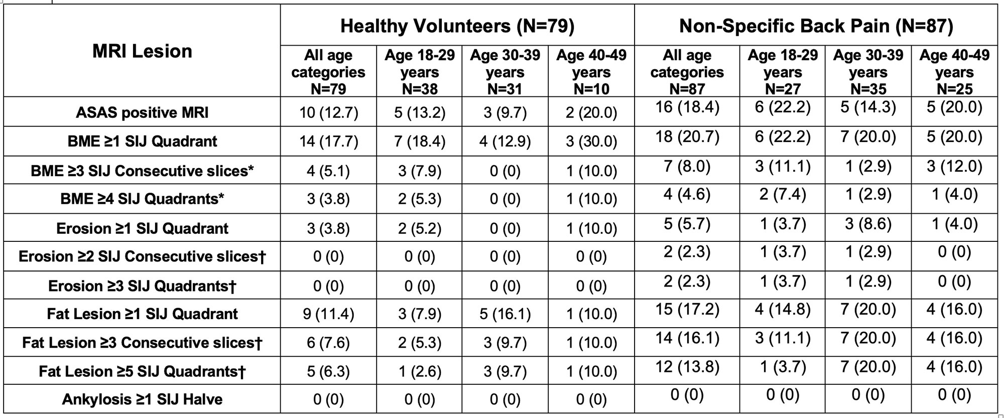
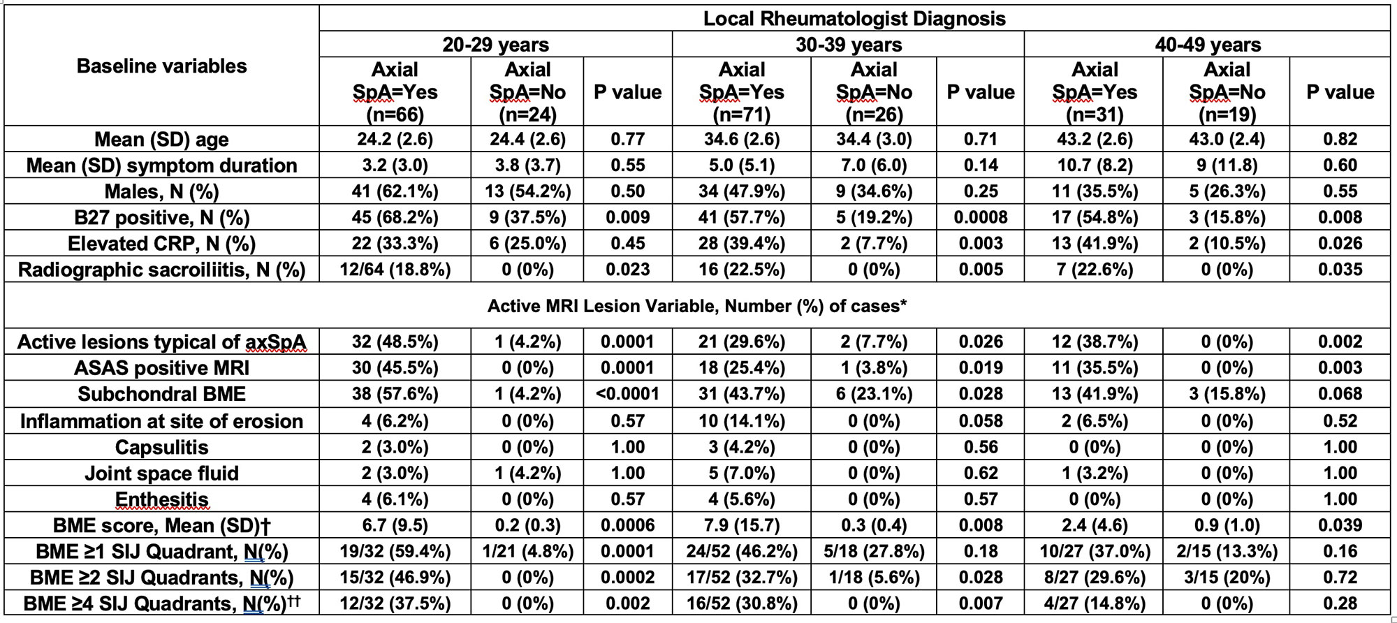
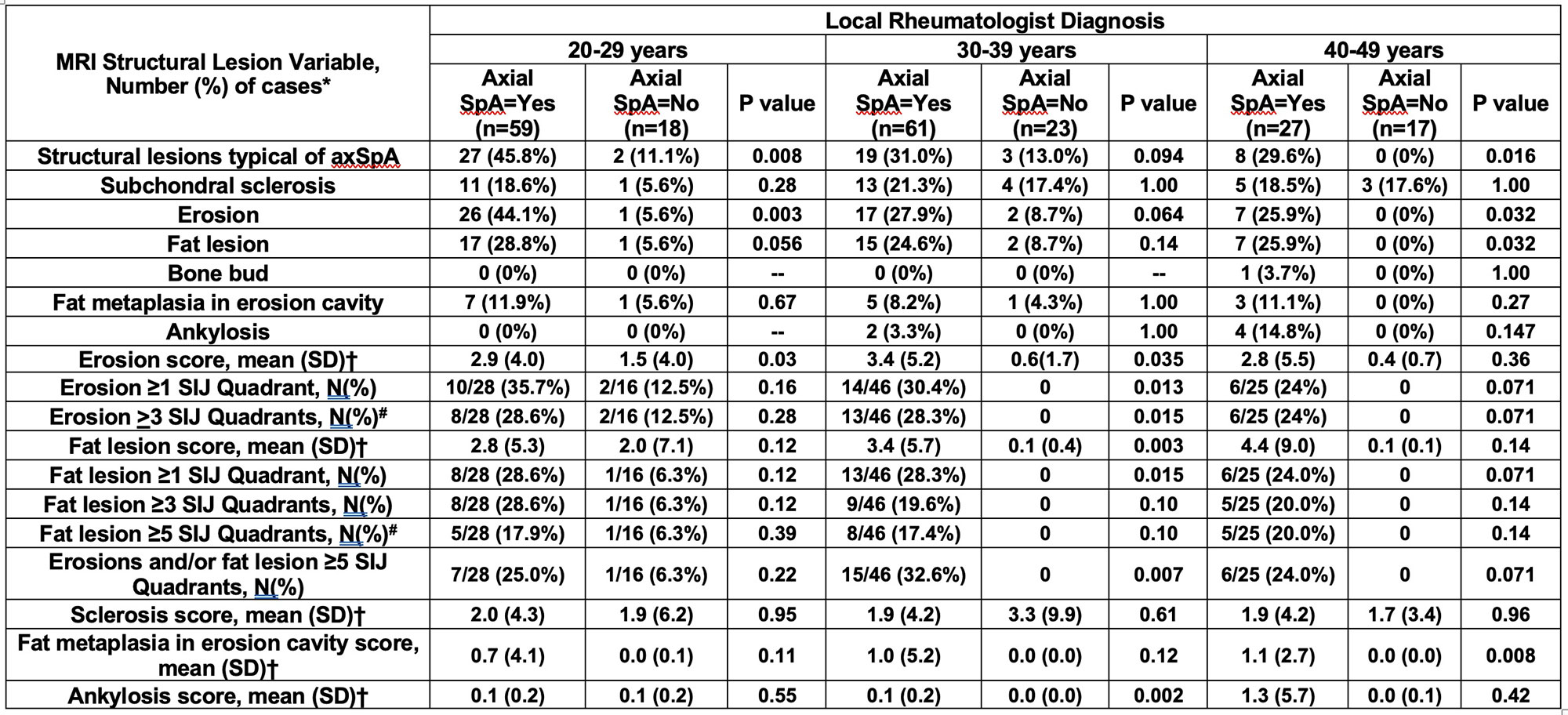
Methods: We assessed MRI SIJ lesions in 2 independent cohorts: A. 79 healthy volunteers (HV) and 87 with NSBP aged ≤49 years from the staffs of two university hospitals in Canada and Switzerland, the absence of back pain being confirmed using the Nordic Questionnaire, B. 237 patients ≤45 years referred to a rheumatologist with undiagnosed back pain suspicious of axSpA in the ASAS Classification Cohort (ASAS-CC), of whom 69 were diagnosed with NSBP. MRI lesions were assessed on an online interface and recorded in SIJ quadrants or halves of consecutive semicoronal slices through the SIJ. Lesions were ascertained by 5 central readers for cohort A and 7 central readers for cohort B according to standardized lesion definitions (CANDEN for cohort A and ASAS definitions for cohort B) and the results analyzed according to age categories and recently reported, data-driven, MRI cut-offs for a positive MRI indicative of inflammatory or structural lesions typical of axSpA (SIJ+/MRIstructure or SIJ+/MRIinflammation)2.

Results: In cohort A (Table 1), the frequencies of BME lesions were higher for HV in the oldest age category (40-49 years), although a progressive increase for NSBP with increasing age was not evident. Only 3/79 (3.8%) of HV and 4/87 (4.6%) of NSBP had BME in ≥4 SIJ quadrants, the cut-off denoting SIJ+/MRIinflammation. Only 3/79 (3.8%) of HV and 5/87 (5.7%) of NSBP had an erosion in ≥1 SIJ quadrant by majority of readers. None of HV and only 2/87 (2.3%) of NSBP had erosion in ≥2 consecutive slices and/or ≥3 SIJ quadrants, the cut-offs denoting SIJ+/MRIstructure. More fat lesions were noted in NSBP than HV but a progressive increase with age was not evident. In cohort B (ASAS-CC), similar frequencies for active and structural lesions were noted in cases with NSBP to cohort A (Tables 2 and 3) and a progressive increase with age was not evident. Moreover, there was not a single case with a SIJ+/MRIstructure in the higher age categories and the finding of erosion was equally discriminatory between axSpA and NSBP, irrespective of age category.

Conclusion: Data from HV and NSBP controls does not indicate progressive increases in MRI structural lesions in the SIJ with increasing age when standardized definitions for reporting axSpA lesions are adopted. MRI cut-offs for a positive MRI indicative of active or structural MRI lesions typical of axSpA discriminate equally well between axSpA and NSBP across age categories.

Ref 1. Renson et al. Arthritis Rheumatol 2022 doi.org/10.1002/art.42145. 2. Maksymowych et al. Rheumatol 2021;60:4778–4789

Supporting image 1

Table 1. Number (%) of Healthy Individuals Without Back Pain and Non-Specific Back Pain Patients with MRI SIJ Lesions According to ASAS Definitions and Majority of Readers. *MRI cut-offs for positive MRI indicative of active lesion typical of axSpA †MRI cut-offs for positive MRI indicative of structural lesion typical of axSpA

Supporting image 2

Table 2. Frequencies of active MRI lesions in the SIJ Recorded by 7 ASAS-MRI Central Readers According to Local Rheumatologist Diagnosis of AxSpA or NSBP in Consecutive Patients presenting with Undiagnosed Back Pain in the ASAS Classification Cohort. *Based on majority reader (≥4/7) agreement †Based on mean of 7 readers ††MRI cut-off for positive MRI indicative of active lesion typical of axSpA

Supporting image 3

Table 3. Frequencies of structural MRI lesions in the SIJ Recorded by 7 ASAS-MRI Central Readers According to Local Rheumatologist Diagnosis of AxSpA or NSBP in Consecutive Patients presenting with Undiagnosed Back Pain in the ASAS Classification Cohort. *Based on majority reader (≥4/7) agreement. †Based on mean of 7 readers #MRI cut-offs for positive MRI indicative of structural lesion typical of axSpA


Disclosures: U. Weber, None; S. Pedersen, Novartis, AbbVie/Abbott, UCB, Pfizer, Merck/MSD; M. Østergaard, AbbVie/Abbott, Amgen, Bristol-Myers Squibb(BMS), Celgene, Eli Lilly, Janssen, Gilead, Novartis, Pfizer, UCB; P. Machado, AbbVie/Abbott, Eli Lilly, UCB, Novartis, Orphazyme, Galapagos; X. Baraliakos, AbbVie, Lilly, Galapagos, MSD, Novartis, Pfizer, UCB, Bristol-Myers Squibb, Janssen, Roche, Sandoz, Sanofi; R. Lambert, Calyx, CARE Arthritis, Image Analysis Group; W. Maksymowych, AbbVie, Boehringer-Ingelheim, Celgene, Eli Lilly, Galapagos, Janssen, Novartis, Pfizer, UCB, CARE Arthritis Limited.

To cite this abstract in AMA style:

Weber U, Pedersen S, Østergaard M, Machado P, Baraliakos X, Lambert R, Maksymowych W. Relative Frequencies of the Spectrum of MRI Lesions in the Sacroiliac Joints of Healthy Individuals and Patients with Non-Specific Back Pain: What Is the Impact of Increasing Age? [abstract]. Arthritis Rheumatol. 2022; 74 (suppl 9). https://acrabstracts.org/abstract/relative-frequencies-of-the-spectrum-of-mri-lesions-in-the-sacroiliac-joints-of-healthy-individuals-and-patients-with-non-specific-back-pain-what-is-the-impact-of-increasing-age/. Accessed .
  • Tweet
  • Click to email a link to a friend (Opens in new window) Email
  • Click to print (Opens in new window) Print

« Back to ACR Convergence 2022

ACR Meeting Abstracts - https://acrabstracts.org/abstract/relative-frequencies-of-the-spectrum-of-mri-lesions-in-the-sacroiliac-joints-of-healthy-individuals-and-patients-with-non-specific-back-pain-what-is-the-impact-of-increasing-age/

Advanced Search

Your Favorites

You can save and print a list of your favorite abstracts during your browser session by clicking the “Favorite” button at the bottom of any abstract. View your favorites »

All abstracts accepted to ACR Convergence are under media embargo once the ACR has notified presenters of their abstract’s acceptance. They may be presented at other meetings or published as manuscripts after this time but should not be discussed in non-scholarly venues or outlets. The following embargo policies are strictly enforced by the ACR.

Accepted abstracts are made available to the public online in advance of the meeting and are published in a special online supplement of our scientific journal, Arthritis & Rheumatology. Information contained in those abstracts may not be released until the abstracts appear online. In an exception to the media embargo, academic institutions, private organizations, and companies with products whose value may be influenced by information contained in an abstract may issue a press release to coincide with the availability of an ACR abstract on the ACR website. However, the ACR continues to require that information that goes beyond that contained in the abstract (e.g., discussion of the abstract done as part of editorial news coverage) is under media embargo until 10:00 AM ET on November 14, 2024. Journalists with access to embargoed information cannot release articles or editorial news coverage before this time. Editorial news coverage is considered original articles/videos developed by employed journalists to report facts, commentary, and subject matter expert quotes in a narrative form using a variety of sources (e.g., research, announcements, press releases, events, etc.).

Violation of this policy may result in the abstract being withdrawn from the meeting and other measures deemed appropriate. Authors are responsible for notifying colleagues, institutions, communications firms, and all other stakeholders related to the development or promotion of the abstract about this policy. If you have questions about the ACR abstract embargo policy, please contact ACR abstracts staff at [email protected].

Wiley

  • Online Journal
  • Privacy Policy
  • Permissions Policies
  • Cookie Preferences

© Copyright 2025 American College of Rheumatology