ACR Meeting Abstracts

ACR Meeting Abstracts

  • Meetings
    • ACR Convergence 2024
    • ACR Convergence 2023
    • 2023 ACR/ARP PRSYM
    • ACR Convergence 2022
    • ACR Convergence 2021
    • ACR Convergence 2020
    • 2020 ACR/ARP PRSYM
    • 2019 ACR/ARP Annual Meeting
    • 2018-2009 Meetings
    • Download Abstracts
  • Keyword Index
  • Advanced Search
  • Your Favorites
    • Favorites
    • Login
    • View and print all favorites
    • Clear all your favorites
  • ACR Meetings

Abstract Number: 2897

Increased CD69+ Tissue-resident Memory T (TRM) Cells and STAT3 Expression in Cutaneous Lupus Erythematosus Patients Recalcitrant to Antimalarials

Majid Zeidi 1, Kristen Chen 1, Krisha Desai 1, Hee Joo Kim 2, Srita Chakka 3, Rachel Lim 4 and Victoria Werth1, 1Corporal Michael J. Crescenz VAMC, Philadelphia, PA, USA and Department of Dermatology, University of Pennsylvania, Philadelphia, PA, USA, Philadelphia, PA, 2Corporal Michael J. Crescenz VAMC, Philadelphia, PA, USA andDepartment of Dermatology, University of Pennsylvania, Philadelphia, PA, USA and Department of Dermatology, Gil Medical Center, Gachon University College of Medicine, Incheon, Korea, Republic of (South), Philadelphia, 3Department of Dermatology, University of Pennsylvania, Philadelphia, PA, USA, Philadelphia, 4Corporal Michael J. Crescenz VAMC, Philadelphia, PA, USA, Philadelphia

Meeting: 2019 ACR/ARP Annual Meeting

Keywords: Antimalarial drugs, cutaneous lupus, dendritic cells and Macrophage, T cells

  • Tweet
  • Click to email a link to a friend (Opens in new window) Email
  • Click to print (Opens in new window) Print
Session Information

Date: Wednesday, November 13, 2019

Title: 6W022: SLE – Etiology & Pathogenesis II (2894–2899)

Session Type: ACR Abstract Session

Session Time: 11:00AM-12:30PM

Background/Purpose: Cutaneous lupus erythematosus (CLE) is an autoimmune disease with various subsets and clinical manifestations. T lymphocytes are the predominant cell type found in lesional skin, but plasmacytoid dendritic cells (pDCs) and myeloid dendritic cells (mDCs) also play an important role in the pathogenesis. Oral antimalarials, including hydroxychloroquine (HCQ) and quinacrine (QC) are first-line systemic therapy for all CLE sub-types. Although HCQ is effective in approximately 50% of patients, some patients still do not respond to antimalarials. Some of these patients benefit from additional QC, but there is a subset of patients who remain refractory to both antimalarials. Refractoriness poses a huge challenge as patients continue to have active disease despite antimalarial therapy. To better understand reasons for refractoriness in CLE, we investigated the immunologic characteristics of patients who responded to antimalarials versus those who did not.

Methods: One hundred fifteen patients with a diagnosis of CLE were recruited from the Autoimmune Skin Disease Center at the Hospital of the University of Pennsylvania. Sixty-five were well-characterized in terms of response to treatment as (i) HCQ-responders (n=22), (ii) HCQ+QC-responders (n=24), or (iii) HCQ+QC-nonresponders (n=19). Lesional skin was biopsied before starting treatment with antimalarials. We defined treatment failure to HCQ as continued skin activity requiring a second intervention after at least 2 months of HCQ therapy. Immunohistochemistry was used to characterize the inflammatory cells and cytokine expression in lesional skin biopsies from patients. Total RNA was extracted from these biopsies to analyze specific gene signatures. The patient’s CLASI score – a measure of disease activity – at the time of the biopsy was also determined.

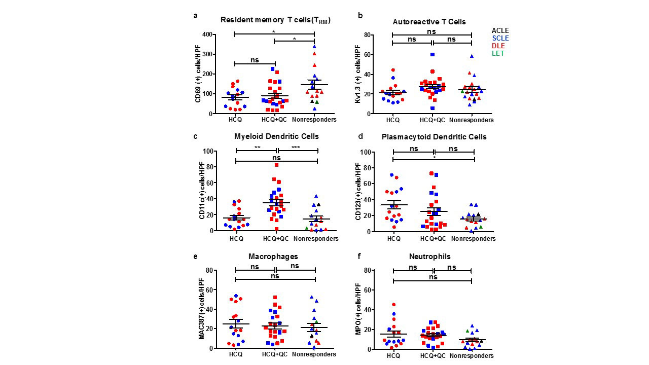
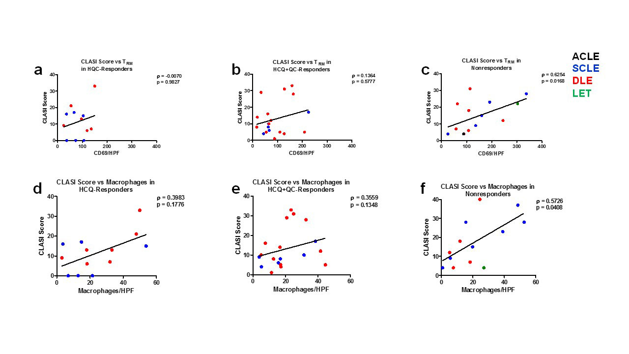
Results: Immunohistochemistry showed that CD69+ tissue-resident memory T (TRM) cells were significantly higher in HCQ+QC-nonresponders compared to HCQ- and HCQ+QC-responders. mDCs were significantly higher in HCQ+QC-responders compared to HCQ- and HCQ+QC-nonresponders. There were significantly higher pDCs in the HCQ-responders compared to the nonresponders. There was no significant difference in the number of autoreactive T cells, macrophages, and neutrophils among the three groups (Figure 1). The HCQ+QC-nonresponder group was distinct from the other groups in that their CLASI scores correlated positively with the number of TRM cells (r=0.6254, p=0.017) and macrophages (r=0.5726, p=0.041) (Figure 2). mRNA expression demonstrated high STAT3 expression in HCQ+QC-nonresponders (Figure 3). There was a significantly higher percentage of area stained for IL-17 in the HCQ+QC-responders compared to HCQ-responders and HCQ+QC-nonresponders while IL-22 expression was not significantly different between groups.

Conclusion: An increased number of CD69+ TRM cells and correlation between CD69+TRM cells and macrophages with CLASI scores in the HCQ+QC-nonresponders, a finding not seen in either HCQ or HCQ+QC-responders, may indicate that CD69+ TRM cells and macrophages are involved in antimalarial-refractory skin disease.

Figure 1: Immune Cell Quantification in HCQ-responders, HCQ+QC-responders, and Nonresponders. There was a significantly higher number of tissue-resident memory T -TRM- cells in nonresponders compared to HCQ-responders and HCQ+QC-responders -p<0.05, Figure 1a-. There was a significantly higher number of mDCs in HCQ+QC-responders compared to the nonresponders -p<0.001, Figure 1c- and HCQ-responders -p<0.01, Figure 1c-. There was a significantly higher number of pDCs in the HCQ-responders compared to the nonresponders -p<0.05, Figure 1d-. There was no significant difference in the number of auto reactive T cells -Figure 1b- macrophages -Figure 1e- and number of neutrophils -Figure 1f- among the three groups.

Figure 2: CLASI Score versus tissue-resident memory T -TRM- cells and Macrophages in HCQ-responders, HCQ+QC-responders, and Nonresponders. For the nonresponders, there was a significant positive correlation between the CLASI score with the number of tissue-resident memory T -TRM- cells and macrophages -p<0.05, Figures 3c&3f-. There was no significant correlation between the CLASI score with the number of tissue-resident memory T -TRM- cells and macrophages counts in the HCQ-responders and HCQ+QC-responders.

Figure 3: Type 1 Interferon, TNF, and STAT Gene Expression in HCQ-responders, HCQ+QC-responders, and Nonresponders. The nonresponders were found to have a significantly decreased type 1 IFN signature and elevated TNF-α expression compared to the HCQ-responders -OAS1 p<0.01; OASL p<0.05; ISG15 p<0.01; LY6E p<0.01; MX1 p<0.05; TNF p<0.01, Figures 4a-f-. In terms of the STAT genes, the nonresponders had increased expression of STAT3 -p<0.05, Figure 4h- and decreased expression of STAT1 -p<0.01, Figure 4g- and STAT4 -p<0.05, Figure 4i- compared to the HCQ-responders.
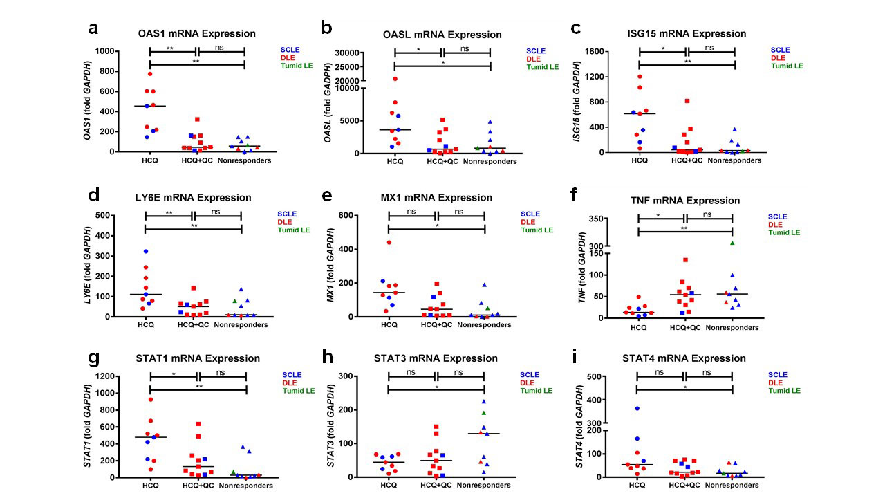
The nonresponders and HCQ+QC-responders had a similar expression profile for all analyzed genes.


Disclosure: M. Zeidi, None; K. Chen, None; K. Desai, None; H. Kim, None; S. Chakka, None; R. Lim, None; V. Werth, Biogen, 2, 5, Corbus Pharmaceuticals, 2, 9, University of Pennsylvania, 9.

To cite this abstract in AMA style:

Zeidi M, Chen K, Desai K, Kim H, Chakka S, Lim R, Werth V. Increased CD69+ Tissue-resident Memory T (TRM) Cells and STAT3 Expression in Cutaneous Lupus Erythematosus Patients Recalcitrant to Antimalarials [abstract]. Arthritis Rheumatol. 2019; 71 (suppl 10). https://acrabstracts.org/abstract/increased-cd69-tissue-resident-memory-t-trm-cells-and-stat3-expression-in-cutaneous-lupus-erythematosus-patients-recalcitrant-to-antimalarials/. Accessed .
  • Tweet
  • Click to email a link to a friend (Opens in new window) Email
  • Click to print (Opens in new window) Print

« Back to 2019 ACR/ARP Annual Meeting

ACR Meeting Abstracts - https://acrabstracts.org/abstract/increased-cd69-tissue-resident-memory-t-trm-cells-and-stat3-expression-in-cutaneous-lupus-erythematosus-patients-recalcitrant-to-antimalarials/

Advanced Search

Your Favorites

You can save and print a list of your favorite abstracts during your browser session by clicking the “Favorite” button at the bottom of any abstract. View your favorites »

All abstracts accepted to ACR Convergence are under media embargo once the ACR has notified presenters of their abstract’s acceptance. They may be presented at other meetings or published as manuscripts after this time but should not be discussed in non-scholarly venues or outlets. The following embargo policies are strictly enforced by the ACR.

Accepted abstracts are made available to the public online in advance of the meeting and are published in a special online supplement of our scientific journal, Arthritis & Rheumatology. Information contained in those abstracts may not be released until the abstracts appear online. In an exception to the media embargo, academic institutions, private organizations, and companies with products whose value may be influenced by information contained in an abstract may issue a press release to coincide with the availability of an ACR abstract on the ACR website. However, the ACR continues to require that information that goes beyond that contained in the abstract (e.g., discussion of the abstract done as part of editorial news coverage) is under media embargo until 10:00 AM ET on November 14, 2024. Journalists with access to embargoed information cannot release articles or editorial news coverage before this time. Editorial news coverage is considered original articles/videos developed by employed journalists to report facts, commentary, and subject matter expert quotes in a narrative form using a variety of sources (e.g., research, announcements, press releases, events, etc.).

Violation of this policy may result in the abstract being withdrawn from the meeting and other measures deemed appropriate. Authors are responsible for notifying colleagues, institutions, communications firms, and all other stakeholders related to the development or promotion of the abstract about this policy. If you have questions about the ACR abstract embargo policy, please contact ACR abstracts staff at [email protected].

Wiley

  • Online Journal
  • Privacy Policy
  • Permissions Policies
  • Cookie Preferences

© Copyright 2025 American College of Rheumatology