ACR Meeting Abstracts

ACR Meeting Abstracts

  • Meetings
    • ACR Convergence 2024
    • ACR Convergence 2023
    • 2023 ACR/ARP PRSYM
    • ACR Convergence 2022
    • ACR Convergence 2021
    • ACR Convergence 2020
    • 2020 ACR/ARP PRSYM
    • 2019 ACR/ARP Annual Meeting
    • 2018-2009 Meetings
    • Download Abstracts
  • Keyword Index
  • Advanced Search
  • Your Favorites
    • Favorites
    • Login
    • View and print all favorites
    • Clear all your favorites
  • ACR Meetings

Abstract Number: 1866

Gene Expression Meta-Analysis Reveals Commonalities in Gene Activation and Enrichment of Immune Pathways and Cell Types in Dermatomyositis Target Tissues

Jessica Neely1, Dmitry Rychkov 1, Manish Paranjpe 1, Michael Waterfield 2, Susan Kim 1 and Marina Sirota 2, 1University of California, San Francisco, San Francisco, CA, 2University of California, San Francisco, San Francisco

Meeting: 2019 ACR/ARP Annual Meeting

Keywords: dermatomyositis, Gene Expression, interferons, juvenile dermatomyositis and genomics

  • Tweet
  • Click to email a link to a friend (Opens in new window) Email
  • Click to print (Opens in new window) Print
Session Information

Date: Monday, November 11, 2019

Title: 4M113: Muscle Biology, Myositis & Myopathies I (1866–1871)

Session Type: ACR Abstract Session

Session Time: 4:30PM-6:00PM

Background/Purpose:

Dermatomyositis (DM) is a complex immune-mediated disease resulting in muscle and skin inflammation.  Prior studies of gene expression in DM have revealed a type I interferon (IFN) signature, but less is known about alternative immune pathways involved.  We conducted a comprehensive gene expression meta-analysis in DM muscle and skin to identify disease-relevant genes and pathways and elucidate a shared immune mechanism across tissues.

Methods: Raw data from 6 publicly available datasets from DM muscle (71 cases, 36 controls) and 2 from skin (77 cases, 22 controls) were downloaded.  Data were preprocessed, merged and batch-corrected, creating tissue-specific gene expression matrices.  Significance Analysis of Microarrays was used to identify differentially expressed genes (DEG; cutoff FDR p-value < 0.05, FC≧1.3).  A hypergeometric test was used to assess overlap between DEGs in skin and muscle.  Weighted Gene Coexpression Network Analysis was used to construct muscle and skin networks and identify associations not detected on the single gene level.  Disease-associated modules were identified by correlating module eigengene with case/control status.  Network preservation was assessed by calculating gene overlap between muscle and skin modules using a hypergeometric test.  Hub genes (gene significance >0.2 & module membership >0.8) in each network were identified.  Cell type enrichment was performed using xCell.

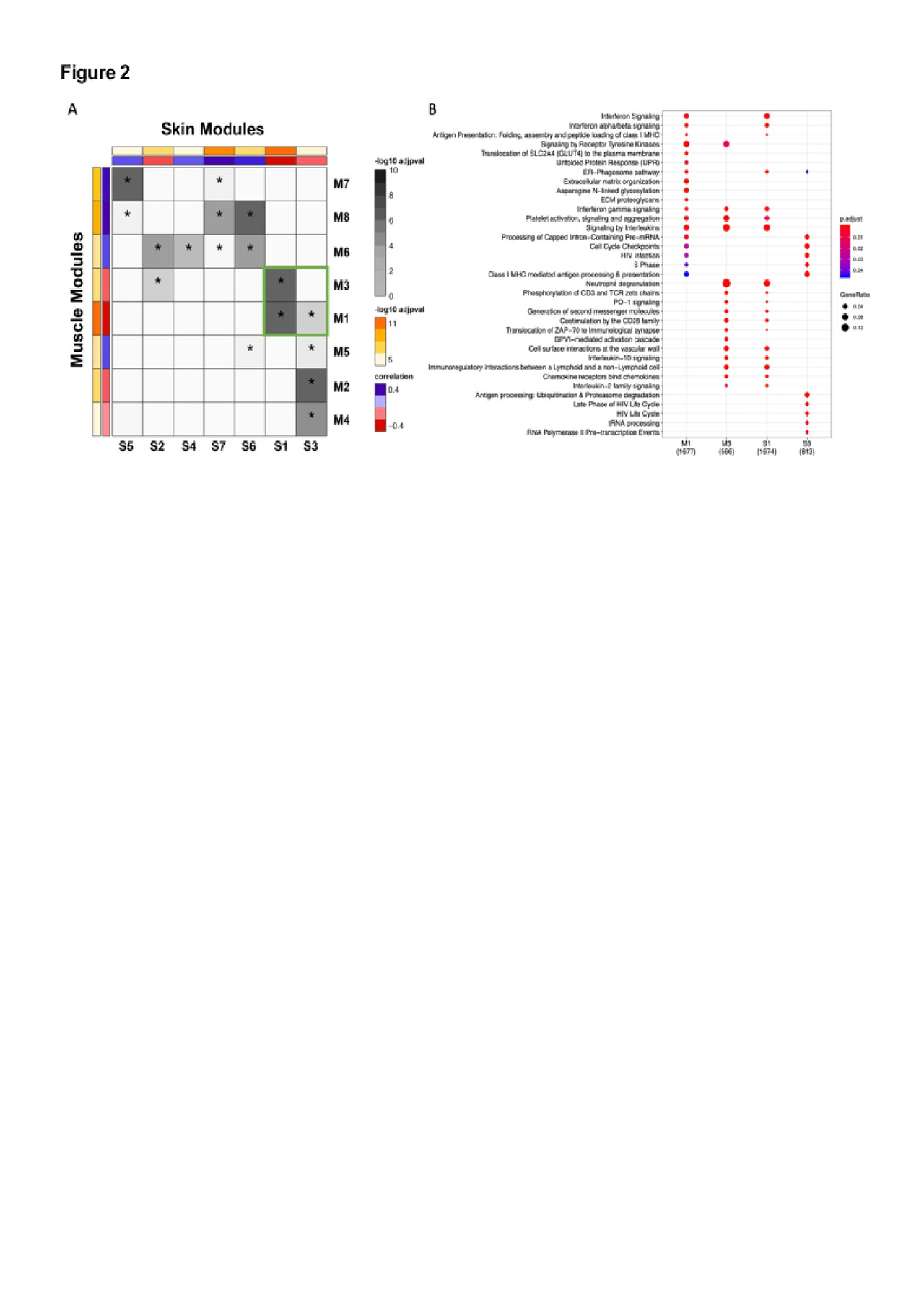
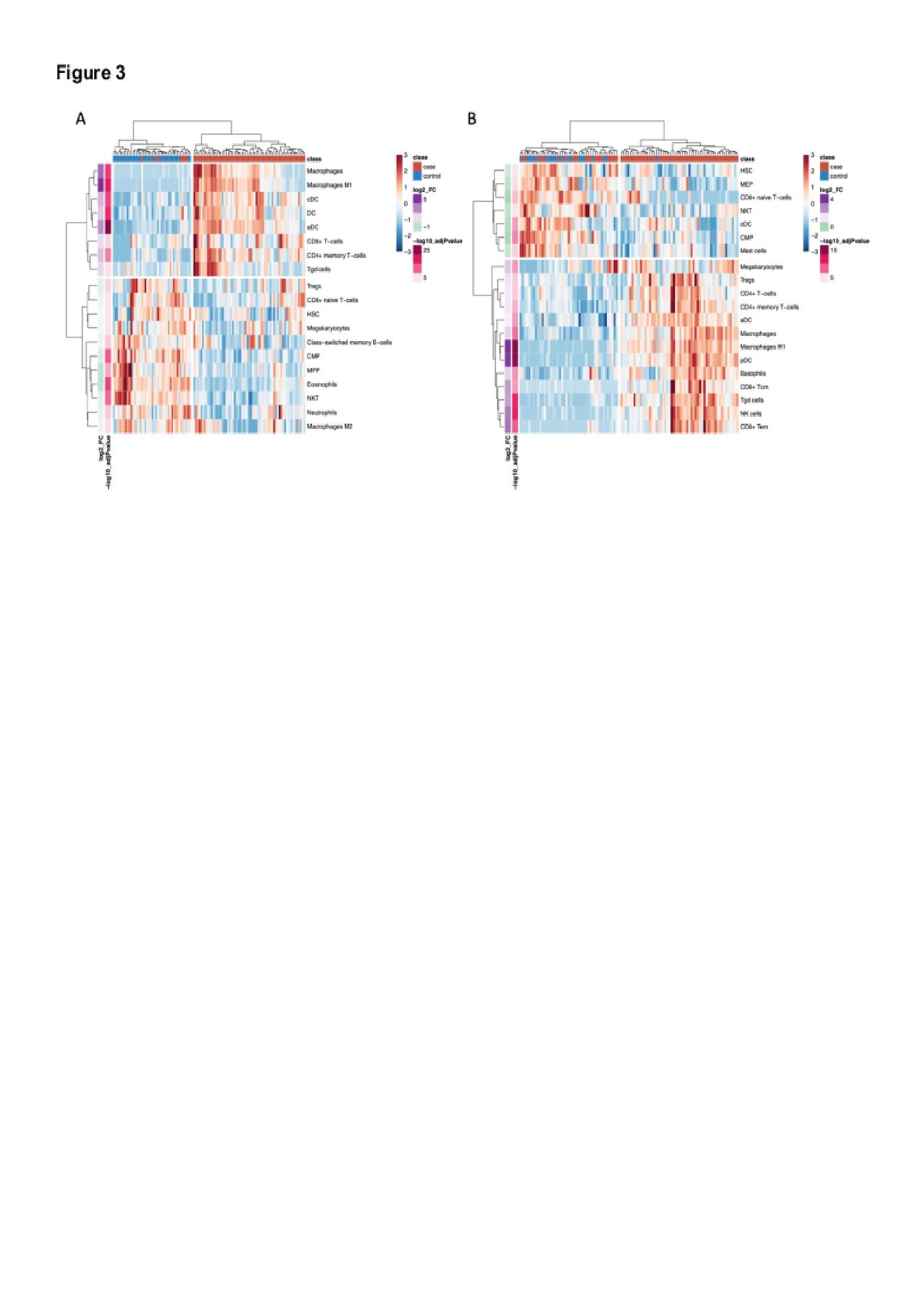
Results: There were 544 significantly DEGs (FC≧1.3, q< 0.05) in muscle and 300 in skin (Fig 1A & 1B).  There was significant overlap in upregulated genes across tissues with 94 shared genes (p=6.4xE-78) which were enriched in type I and II IFN signaling and MHC class I antigen processing pathways (Fig 1C & 1D).  In a network analysis, we identified 8 significant muscle modules and 7 skin modules either positively or negatively correlated with case/control status (Fig 2A).  Modules that were highly correlated with cases also shared significant overlap on the gene level (Fig 2A).  These modules were enriched in pathways consistent with the single gene analysis and also in terms related to T cell activation and T cell receptor signaling (Fig 2B).  Modules correlated with controls were enriched in processes related to energy metabolism.  The top hub genes in each network included genes classically involved in type I IFN (STAT1, MX1, IFI44, ISG15), type II IFN (CXCL10, GBP1), class I MHC antigen processing (HLA-A, -B, -F, B2M, TAP1) and the immunoproteasome (PSMB8, PSMB9).  In the cell type enrichment analysis, both tissues were enriched in antigen-presenting cells, including activated dendritic cells and M1 macrophages (Fig 3A & 3B).

Conclusion:

This analysis demonstrates striking similarities in gene expression in target tissues in DM and simultaneous activation of innate and adaptive immune responses.  Using an unbiased approach, we show enrichment of type I and type II IFN pathways, MHC class I antigen processing pathways, T cell activation, and antigen-presenting cells.  These results suggest type II IFN contributes to the global IFN signature in DM and that altered autoantigen presentation through the class I MHC pathway may be important in disease pathogenesis.


ACR gene expr Fig 1

Heatmaps of differentially expressed genes -DEG- identified by SAM in muscle -A- and skin -B- using cut off of FDR p-value <0.05 and FC 1.3. Hierarchical clustering demonstrates clustering of cases and controls and that the majority of DEGs in both tissues are up-regulated. C- Gene symbols of 94 overlapping differentially expressed genes plotted by fold change in muscle and skin. D- Enrichment of 94 overlapping genes by over-representation analysis using the Reactome Database. P-values are calculated using a hypergeometric distribution and corrected for multiple comparisons by the Benjamini Hochberg method.


ACR gene expr Figure 2

A- Heatmap demonstrating hierarchical clustering of significant modules identified by WGCNA in each tissue based on gene overlap. Correlation between module eigengene and disease status is shown by the red to blue bar where red represents modules most positively-correlated with cases and blue represents modules negatively-correlated with cases. The color corresponds to a signed p-value: sign-correlation- * -log10-FDR p value-. The -log10 adjusted p-value of this correlation is indicated by the yellow to orange bar. The degree of pairwise overlap in genes between muscle modules and skin modules computed using a hypergeometric test is denoted by grey coloring where modules with significant overlap in genes -p <0.05- are denoted by an asterisk and the color range corresponds to –log10-pvalue- of the pairwise overlap. B- Plot showing over-representation analysis of pathways from the Reactome database across a cluster of highly overlapping and interesting modules -green box in pane A- in each tissue demonstrating enrichment of shared pathways across tissues.


ACR gene expr Figure 3

Heatmaps of cell-type enrichment calculated using xCell demonstrates clustering of cases and controls based on cell types in both muscle -A- and skin -B-. Enrichment was strongest for dendritic cells and M1 macrophages in both tissues.


Disclosure: J. Neely, None; D. Rychkov, None; M. Paranjpe, None; M. Waterfield, None; S. Kim, None; M. Sirota, None.

To cite this abstract in AMA style:

Neely J, Rychkov D, Paranjpe M, Waterfield M, Kim S, Sirota M. Gene Expression Meta-Analysis Reveals Commonalities in Gene Activation and Enrichment of Immune Pathways and Cell Types in Dermatomyositis Target Tissues [abstract]. Arthritis Rheumatol. 2019; 71 (suppl 10). https://acrabstracts.org/abstract/gene-expression-meta-analysis-reveals-commonalities-in-gene-activation-and-enrichment-of-immune-pathways-and-cell-types-in-dermatomyositis-target-tissues/. Accessed .
  • Tweet
  • Click to email a link to a friend (Opens in new window) Email
  • Click to print (Opens in new window) Print

« Back to 2019 ACR/ARP Annual Meeting

ACR Meeting Abstracts - https://acrabstracts.org/abstract/gene-expression-meta-analysis-reveals-commonalities-in-gene-activation-and-enrichment-of-immune-pathways-and-cell-types-in-dermatomyositis-target-tissues/

Advanced Search

Your Favorites

You can save and print a list of your favorite abstracts during your browser session by clicking the “Favorite” button at the bottom of any abstract. View your favorites »

All abstracts accepted to ACR Convergence are under media embargo once the ACR has notified presenters of their abstract’s acceptance. They may be presented at other meetings or published as manuscripts after this time but should not be discussed in non-scholarly venues or outlets. The following embargo policies are strictly enforced by the ACR.

Accepted abstracts are made available to the public online in advance of the meeting and are published in a special online supplement of our scientific journal, Arthritis & Rheumatology. Information contained in those abstracts may not be released until the abstracts appear online. In an exception to the media embargo, academic institutions, private organizations, and companies with products whose value may be influenced by information contained in an abstract may issue a press release to coincide with the availability of an ACR abstract on the ACR website. However, the ACR continues to require that information that goes beyond that contained in the abstract (e.g., discussion of the abstract done as part of editorial news coverage) is under media embargo until 10:00 AM ET on November 14, 2024. Journalists with access to embargoed information cannot release articles or editorial news coverage before this time. Editorial news coverage is considered original articles/videos developed by employed journalists to report facts, commentary, and subject matter expert quotes in a narrative form using a variety of sources (e.g., research, announcements, press releases, events, etc.).

Violation of this policy may result in the abstract being withdrawn from the meeting and other measures deemed appropriate. Authors are responsible for notifying colleagues, institutions, communications firms, and all other stakeholders related to the development or promotion of the abstract about this policy. If you have questions about the ACR abstract embargo policy, please contact ACR abstracts staff at [email protected].

Wiley

  • Online Journal
  • Privacy Policy
  • Permissions Policies
  • Cookie Preferences

© Copyright 2025 American College of Rheumatology