Session Information
Date: Monday, October 22, 2018
Title: 4M107 ACR Abstract: SLE–Clinical II: Renal & Neuropsychiatric Disease in SLE (1941–1945)
Session Type: ACR Concurrent Abstract Session
Session Time: 4:30PM-6:00PM
Background/Purpose: Cognitive impairment is common in SLE and impacts quality of life; however, causality is limited by incomplete understanding of neurotoxic mechanisms. Diffusion tensor imaging (DTI) is an advanced MRI technique that assesses white matter integrity (WMI). In an animal model we demonstrated cross-reactive anti-dsDNA/NMDA receptor antibodies (DNRAb) that cause neuron loss, hippocampal dysfunction, and an established behavioral phenotype. In SLE patients, serum DNRAb correlates with poor spatial memory and hippocampal atrophy. We report new associations among serum DNRAb, regional microstructural integrity (DTI) and spatial memory in SLE.
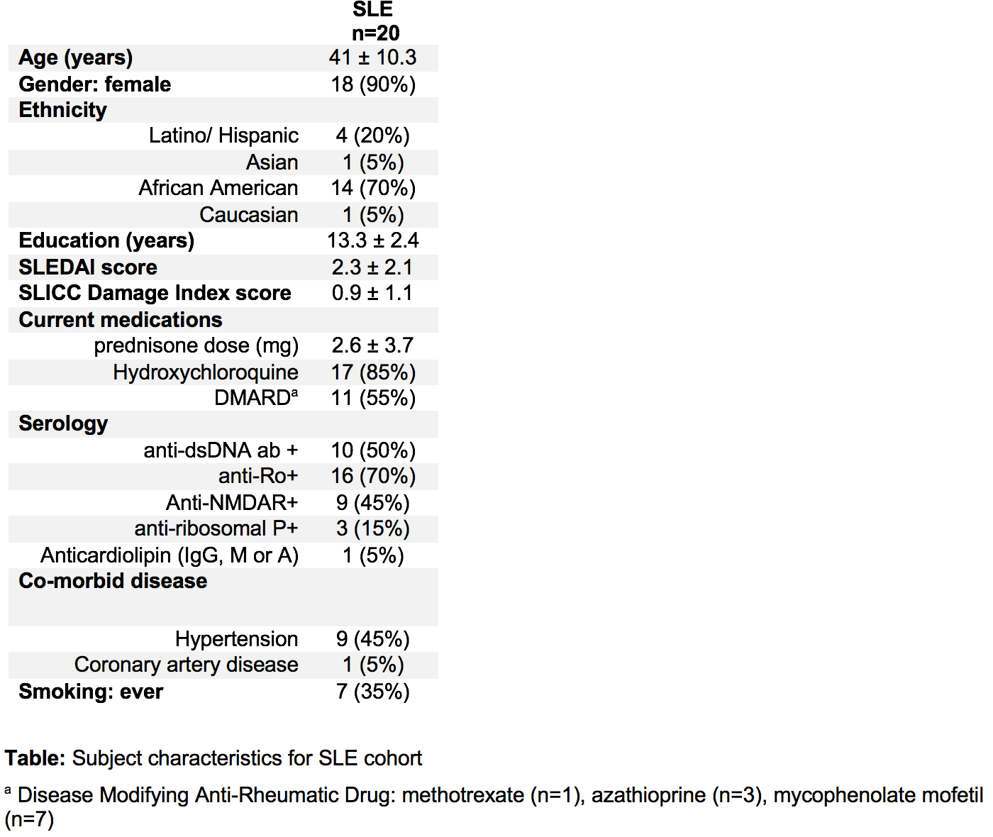
Methods: 19 out of a cohort of 20 adult SLE subjects that met ACR criteria and had no history of CNS insult, well-controlled hypertension, low disease activity and corticosteroid doses (Table), and 14 age/gender matched healthy controls (HC) underwent DTI. A spatial memory task (SMT) assessed both non-spatial object recognition and memory for spatial relations in SLE subjects. Serum DNRAb assays were performed by ELISA with the DWEYS consensus sequence. Fractional anisotropy (FA) maps were compared between SLE and HC, and tract count differences were evaluated (t-test). Regression analysis (Pearson product-moment) assessed correlations between FA maps, serum DNRAb, and spatial memory performance.
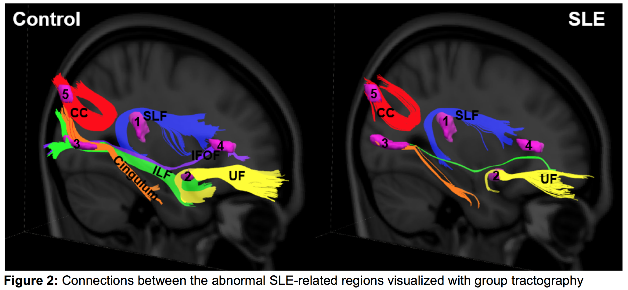
Results: FA was significantly reduced in 5 discrete regions with abnormal WMI in SLE. FA in the parahippocampal gyrus (PHG) correlated inversely with serum DNRAb and positively with SMT performance (Fig. 1). Other regional abnormalities in WM tracts (expressed as % change relative to HC) included: 1) superior longitudinal fasciculus (temporal part) (SLF, -74%), 2) uncinate fasciculus (UF, -86%), 3) cingulum (hippocampus part) (-82%), 4) inferior longitudinal fasciculus (ILF, -99.5%) and inferior frontal occipital fasciculus (IFOF, -100%), and 5) the splenium of the corpus callosum (CC, -48%) (p < 0.001) (Fig. 2).
Conclusion: DTI revealed PHG abnormality that correlated with serum DNRAb and SMT performance, suggesting potential roles of these assessments as biomarkers for cognitive impairment. Other discrete regions of decreased WMI, connected by diminished tracts, raise the specter of clinically unrecognized neurodegeneration in quiescent, stable SLE patients without known CNS disease.
To cite this abstract in AMA style:
Anderson E, Vo A, Ploran E, Diamond B, Volpe B, Aranow C, Eidelberg D, Mackay M. Anti-NMDA Receptor Antibodies in Systemic Lupus Erythematosus Associate with Decreased White Matter Integrity and Impaired Spatial Memory [abstract]. Arthritis Rheumatol. 2018; 70 (suppl 9). https://acrabstracts.org/abstract/anti-nmda-receptor-antibodies-in-systemic-lupus-erythematosus-associate-with-decreased-white-matter-integrity-and-impaired-spatial-memory/. Accessed .« Back to 2018 ACR/ARHP Annual Meeting
ACR Meeting Abstracts - https://acrabstracts.org/abstract/anti-nmda-receptor-antibodies-in-systemic-lupus-erythematosus-associate-with-decreased-white-matter-integrity-and-impaired-spatial-memory/


