Session Information
Date: Sunday, October 21, 2018
Session Type: ACR Poster Session A
Session Time: 9:00AM-11:00AM
Background/Purpose: The objective of the study was to compare magnetic resonance imaging (MRI) and conventional radiography of the sacroiliac joints (SIJs) for detection of structural lesions typical for axial spondyloarthritis (axSpA) in an international multireader exercise with central reading of images in the ASAS cohort.
Methods: Patients with symptoms suggestive of SpA from the international ASAS Cohort who had both radiographs and T1-weighted MRIs of SIJs available for central reading were included in the study. MRI scans were assessed for structural changes compatible with axSpA (global statement) and separate changes such as erosion, sclerosis, fat metaplasia and ankylosis, by 7 central readers. Structural changes were considered as present if recorded by the majority of readers (at least 4 out of 7). Similar to MRIs, radiographs of the SIJs were scored by 3 different central readers according to the grading system of the modified New York (mNY) criteria. At the patient level, presence of definite structural damage on radiographs was defined as fulfillment of the radiographic criterion of the mNY criteria (sacroiliitis of at least grade 2 bilaterally or at least grade 3 unilaterally); at the single joint level, radiographic structural damage was defined as presence of sacroiliitis grade 2 in the opinion of at least 2 readers. Absolute agreement (percentage of patients / joints with or without structural changes on both MRI and radiography) and Kappa coefficient of agreement between MRI and radiography were determined.
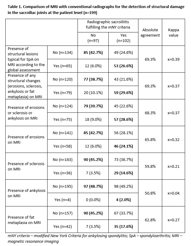
Results: Overall, 199 patients (contributing 398 joints) were included. 149 (74.9%) had a diagnosis of axSpA by a local rheumatologist. Based on central reading, 102 (51.3%) had definite radiographic sacroiliitis according to the mNY Criteria, while 65 (32.7%) had structural changes suggestive of SpA on MRI according to the global assessment. The absolute agreement between MRI and radiographic assessment was 69.3% and kappa was 0.39 (Table 1). Structural lesions scored positive on x-rays could not be confirmed in a relative high percentage (48.1%) on MRI. At the single joint level, the absolute agreement between MRI and radiography was 70.4% and kappa was 0.39 (Table 2). Among structural lesions, erosions on MRI showed the best discriminative capacity regarding the structural damage on radiographs (Tables 1 and 2).
Conclusion: There was only modest agreement between MRI and conventional radiography in terms of detection of structural changes typical for SpA in the SIJs. Erosions on MRI showed the best agreement with the presence of definite structural damage on radiographs.
To cite this abstract in AMA style:
Protopopov M, Poddubnyy D, Proft F, Wichuk S, Machado P, Lambert RG, Weber U, Pedersen SJ, Østergaard M, Sieper J, Rudwaleit M, Baraliakos X, Maksymowych WP. Comparison of MRI and Conventional Radiography for Detection of Structural Changes Typical for Spa – Data from the Assessment of Spondyloarthritis International Society Cohort [abstract]. Arthritis Rheumatol. 2018; 70 (suppl 9). https://acrabstracts.org/abstract/comparison-of-mri-and-conventional-radiography-for-detection-of-structural-changes-typical-for-spa-data-from-the-assessment-of-spondyloarthritis-international-society-cohort/. Accessed .« Back to 2018 ACR/ARHP Annual Meeting
ACR Meeting Abstracts - https://acrabstracts.org/abstract/comparison-of-mri-and-conventional-radiography-for-detection-of-structural-changes-typical-for-spa-data-from-the-assessment-of-spondyloarthritis-international-society-cohort/


