ACR Meeting Abstracts

ACR Meeting Abstracts

  • Meetings
    • ACR Convergence 2024
    • ACR Convergence 2023
    • 2023 ACR/ARP PRSYM
    • ACR Convergence 2022
    • ACR Convergence 2021
    • ACR Convergence 2020
    • 2020 ACR/ARP PRSYM
    • 2019 ACR/ARP Annual Meeting
    • 2018-2009 Meetings
    • Download Abstracts
  • Keyword Index
  • Advanced Search
  • Your Favorites
    • Favorites
    • Login
    • View and print all favorites
    • Clear all your favorites
  • ACR Meetings

Abstract Number: 1997

Detection of Progression of Radiographic Damage Despite Early and Targeted Therapy in Inflammatory Arthritis Using High-Resolution Peripheral Quantitative Computed Tomography

Sarah Manske1,2, Stephanie Finzel3, Steven K. Boyd4 and Cheryl Barnabe5, 1Department of Radiology, University of Calgary, Calgary, AB, Canada, 2McCaig Institute for Bone and Joint Health, University of Calgary, Calgary, AB, Canada, 3Department of Rheumatology and Clinical Immunology, Medical Center University of Freiburg, Faculty of Medicine, University of Freiburg, Freiburg, Germany, 4Cumming School of Medicine, University of Calgary, Calgary, AB, Canada, 5Division of Rheumatology, University of Calgary, Calgary, AB, Canada

Meeting: 2017 ACR/ARHP Annual Meeting

Date of first publication: September 18, 2017

Keywords: Bone, Early Rheumatoid Arthritis, HR-pQCT and treatment

  • Tweet
  • Click to email a link to a friend (Opens in new window) Email
  • Click to print (Opens in new window) Print
Session Information

Date: Tuesday, November 7, 2017

Title: Imaging of Rheumatic Diseases Poster II

Session Type: ACR Poster Session C

Session Time: 9:00AM-11:00AM

Background/Purpose: Early initiation of DMARDs and targeted therapy to achieve remission are said to offer the best opportunity to prevent radiographic damage in early inflammatory arthritis (EIA). High-resolution peripheral quantitative computed tomography (HR-pQCT) provides superior resolution of bone compared to x-ray, and reliable quantification of joint space [1] and erosions [2]. We used HR-pQCT to quantify changes in joint space, erosion number and size, and bone microarchitecture in EIA patients during the first year of therapy.

Methods: We recruited 47 EIA subjects and performed HR-pQCT (XtremeCT I, Scanco Medical AG, Switzerland) scans of the 2nd and 3rd MCPs of their dominant hand at diagnosis and 1 year later and collected sociodemographic and disease characteristics and disease activity at both timepoints. Erosion number and size were assessed from HR-pQCT images by applying the erosion definition and margin landmarks proposed by SPECTRA [2]. Mean, maximum, and minimum joint space width as well as joint space volume were assessed in 3D [1]. Total bone mineral density (Tt.BMD) was assessed in the distal 12% of the 2nd and 3rd metacarpal heads.

Results: At baseline, 34 met classification criteria for RA [3] and 13 had undifferentiated arthritis. After 1 year several individuals were reclassified: 1 had adult onset StillÕs disease, 1 had Psoriatic Arthritis, and 3 had a self-limited arthritis. The majority received DMARD therapy (89%) and 27% progressed to biologic use. The mean DAS28 improved from 5.4 (SD 1.2) to 2.4 (SD 1.1), and the HAQ decreased from 1.20 (SD 0.72) to 0.41 (SD 0.51). Erosions by HR-pQCT were found in 34% (19) of subjects at baseline, with 32 erosions detected (mean axial width 1.2 (SD 0.7) mm, axial depth 1.8 (SD 1.1) mm, perpendicular width 1.4 (SD 0.9) mm, perpendicular depth 1.8 (SD 1.1) mm. This increased to 40 erosions at 1 year, with 23 subjects remaining free of erosions, 12 having a stable number and 8 having an increased number of erosions. However, only 3 erosions had a depth increase that exceeded the smallest detectable change. At the 3rd MCP, mean and minimum joint space width (mm) decreased by 0.06 (SD 0.11) and 0.082 (SD 0.20), respectively. In contrast there were no significant changes in joint space width or volume at the 2nd MCP joint. There were no significant changes in Tt.BMD in the 2nd or 3rd MC. Type of therapy, CCP, RF, or 1-year remission status did not influence change in Tt.BMD or joint space.

Conclusion: Joint space narrowing progressed and the number of erosions increased despite rheumatologic intervention. HR-pQCT provides greater sensitivity for detection of changes in joint space, erosions and bone microarchitecture.

Erosion%20Figure%20v2.png

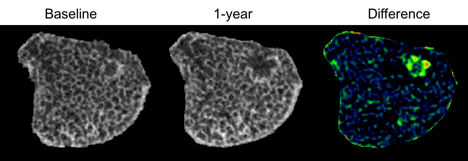
Figure 1. Change in erosion over 1-year follow-up evaluated with HR-pQCT after registration and alignment with baseline image.

References: [1] Barnabe et al, Med Eng Phys, 35: 1540-44, 2013. [2] Barnabe et al, J Rheumatol, 43: 1935-40, 2016. [3] Aletaha et al, Arthitis Rheum, 62: 2569-81, 2010.


Disclosure: S. Manske, None; S. Finzel, None; S. K. Boyd, None; C. Barnabe, None.

To cite this abstract in AMA style:

Manske S, Finzel S, Boyd SK, Barnabe C. Detection of Progression of Radiographic Damage Despite Early and Targeted Therapy in Inflammatory Arthritis Using High-Resolution Peripheral Quantitative Computed Tomography [abstract]. Arthritis Rheumatol. 2017; 69 (suppl 10). https://acrabstracts.org/abstract/detection-of-progression-of-radiographic-damage-despite-early-and-targeted-therapy-in-inflammatory-arthritis-using-high-resolution-peripheral-quantitative-computed-tomography/. Accessed .
  • Tweet
  • Click to email a link to a friend (Opens in new window) Email
  • Click to print (Opens in new window) Print

« Back to 2017 ACR/ARHP Annual Meeting

ACR Meeting Abstracts - https://acrabstracts.org/abstract/detection-of-progression-of-radiographic-damage-despite-early-and-targeted-therapy-in-inflammatory-arthritis-using-high-resolution-peripheral-quantitative-computed-tomography/

Advanced Search

Your Favorites

You can save and print a list of your favorite abstracts during your browser session by clicking the “Favorite” button at the bottom of any abstract. View your favorites »

All abstracts accepted to ACR Convergence are under media embargo once the ACR has notified presenters of their abstract’s acceptance. They may be presented at other meetings or published as manuscripts after this time but should not be discussed in non-scholarly venues or outlets. The following embargo policies are strictly enforced by the ACR.

Accepted abstracts are made available to the public online in advance of the meeting and are published in a special online supplement of our scientific journal, Arthritis & Rheumatology. Information contained in those abstracts may not be released until the abstracts appear online. In an exception to the media embargo, academic institutions, private organizations, and companies with products whose value may be influenced by information contained in an abstract may issue a press release to coincide with the availability of an ACR abstract on the ACR website. However, the ACR continues to require that information that goes beyond that contained in the abstract (e.g., discussion of the abstract done as part of editorial news coverage) is under media embargo until 10:00 AM ET on November 14, 2024. Journalists with access to embargoed information cannot release articles or editorial news coverage before this time. Editorial news coverage is considered original articles/videos developed by employed journalists to report facts, commentary, and subject matter expert quotes in a narrative form using a variety of sources (e.g., research, announcements, press releases, events, etc.).

Violation of this policy may result in the abstract being withdrawn from the meeting and other measures deemed appropriate. Authors are responsible for notifying colleagues, institutions, communications firms, and all other stakeholders related to the development or promotion of the abstract about this policy. If you have questions about the ACR abstract embargo policy, please contact ACR abstracts staff at [email protected].

Wiley

  • Online Journal
  • Privacy Policy
  • Permissions Policies
  • Cookie Preferences

© Copyright 2025 American College of Rheumatology