Session Information
Session Type: ACR Poster Session A
Session Time: 9:00AM-11:00AM
Background/Purpose: Approximately 50% of patients with systemic sclerosis (SSc) will develop painful digital (finger) ulcers (DUs) at some point during their disease course. These lesions can be extremely disabling and are often refractory to treatment, requiring close monitoring of healing progression. Also, DUs are often the primary outcome measure in clinical trials of SSc-related digital vasculopathy. Our aims were to: (1) demonstrate the feasibility of patients with SSc taking smartphone photographs of their DUs, and (2) use software image analysis on collected images to track lesion area as a marker of wound status.
Methods: Patients with SSc and incident DUs were asked to photograph their lesion(s), using their own smartphone, once per day for a maximum period of 35 days. Patients received normal clinical wound care for the duration of the study, which in most cases was patient self-management. Image length scales were initially calibrated using a fixed (non-varying) object in each image sequence, allowing relative length/area tracking throughout a sequence. Using digital planimetry software (developed and successfully tested in previous work on digital ulcer photographs [1]), lesion area was measured by fitting an elliptical shape to the wound image by a single observer. Areas from each image were then normalised to the area measured first in the time sequence.
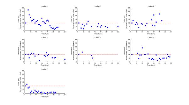
Results: Image sequences describing 7 lesions were collected in-person at the end of the study period from 4 patients. The 7 image sequences cover median [range] duration of 29 [13-35] days. The relative area time course for each lesion is shown in Figure 1. At sequence end, relative lesion areas had, on average, reduced to 56% of the area measured on day 1, with 6 out of 7 lesions reducing in size over the time course.
Conclusion: We have demonstrated the feasibility of patients with SSc collecting images of digital ulcers using smartphone cameras. Images collected are of sufficient quality to allow software monitoring of wound progression (healing or worsening). Further work to build a smartphone app for lesion monitoring (for use in both clinical practice and as an outcome measure in clinical trials) is now required.
References
1. Simpson et al. Arthritis Care & Research, 2017, doi:10.1002/acr.23300
Figure 1. Normalised lesion area time course plots (blue squares) for each of seven digital ulcers. Red dashed line equals 100% relative area, i.e. equal to the value at the start of the sequence. Only lesion 3 showed an increase in area over the course of the study.
To cite this abstract in AMA style:
Dinsdale G, Moore T, Manning J, Murray A, Atkinson R, Ousey K, Dickinson M, Taylor C, Herrick AL. Tracking Digital Ulcers in Systemic Sclerosis: Feasibility Study Assessing Lesion Area from Patient-Recorded Smartphone Photographs [abstract]. Arthritis Rheumatol. 2017; 69 (suppl 10). https://acrabstracts.org/abstract/tracking-digital-ulcers-in-systemic-sclerosis-feasibility-study-assessing-lesion-area-from-patient-recorded-smartphone-photographs/. Accessed .« Back to 2017 ACR/ARHP Annual Meeting
ACR Meeting Abstracts - https://acrabstracts.org/abstract/tracking-digital-ulcers-in-systemic-sclerosis-feasibility-study-assessing-lesion-area-from-patient-recorded-smartphone-photographs/

