Session Information
Session Type: Abstract Submissions (ACR)
Background/Purpose: Several risk factors for osteoarthritis (OA) have been described to be associated with an increased risk for incident radiographic OA, on a local (joint) or systemic (person) level. While radiography depicts articular changes only late in the disease process, magnetic resonance imaging (MRI) is capable of visualizing tissue pathology at a much earlier stage. Most MRI-based studies have used a one knee per person approach and thus data on bilaterality of OA features is sparse. Study aim was to describe symmetricity of MRI-detected OA features in a cohort with knee pain.
Methods: 169 subjects aged 35-65 with chronic, frequent knee pain were included in the Joint in Glucosamine (JOG) study. 3T MRI of both knees was performed using the same pulse sequence protocol as in the Osteoarthritis Initiative (OAI). Knees were semiquantitatively assessed according to the WORMS system by one expert MSK radiologist. Cartilage damage and bone marrow lesions (BMLs) were read in five plates (medial/lateral femur, medial lateral tibia, patella, femoral trochlea) while meniscal damage was read in three medial and three lateral subregions. Chi2 tests were used to compare the proportion of people with unilateral tissue pathology to the proportion what would be expected if the two knees were independent. For this analysis, all MRI features were divided into present (score≥1) and absent (score=0). We further used linear weighted (w) kappa statistics to describe agreement between knees for cartilage damage and BMLs in the same articular plates using the full WORMS scores (0-4 for cartilage and 0-3 for BML).
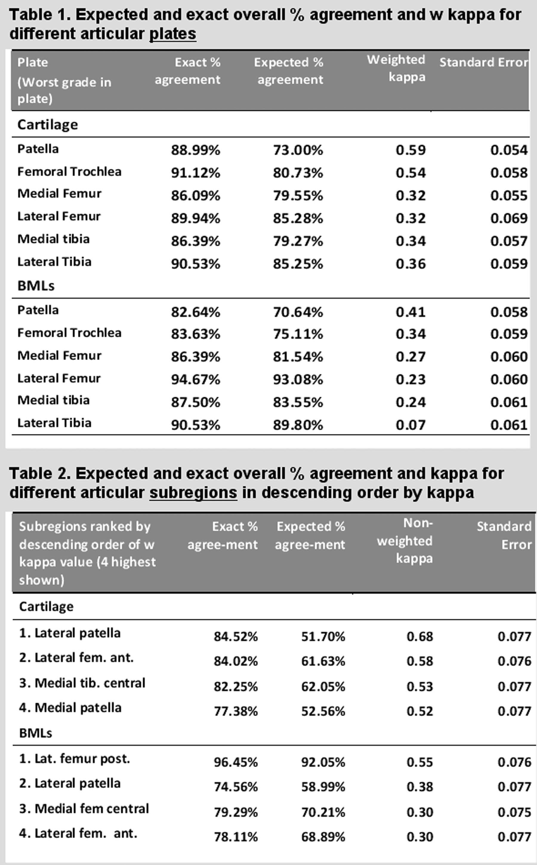
Results: 52.1 % of participants were men, mean age was 51.2 (±6.2) years old, mean BMI was 29.0 (± 4.1). The worst Kellgren/Lawrence (KL) grades in either knee were: K/L 0: 37 (21.9%) knees, K/L 1: 14 (8.3%) knees, K/L 2: 26 (15.4%) knees, K/L 3: 78 (46.2%) knees K/L 4: 14 (8.3). All plates showed a significant lower degree of unilaterality for any cartilage damage (ranging between 15.5% and 32.0%) than expected (ranging between 27.1% and 50.2%). For any BMLs the degree of unilaterality was lower for the patella, trochlea, medial tibia, and medial femur; for any meniscal damage the degree of unilaterality was lower for all medial meniscal subregions but not lateral. All plates showed higher overall % agreement (range 82.6-94.7%) than expected (range 73.0-93.1%) for cartilage damage and BMLs. Moderate agreement (defined as w-kappa 0.4-0.6) was observed for patellar and trochlear cartilage damage (0.59 and 0.54) and patellar (0.41) BMLs.
Conclusion: A higher degree of symmetricity of articular tissue damage than expected by chance was observed in this cohort of subjects with knee pain. These findings support the hypothesis that OA is a multifactorial disease triggered by risk factors on an individual joint level but also by person-based risk factors that predispose joints not only to radiographic OA but also to articular tissue damage commonly associated with OA.
Disclosure:
F. Roemer,
Boston Imaging Core Lab,
1,
National Institute of Health,
5,
Merck Serono,
5;
C. K. Kwoh,
None;
M. J. Hannon,
None;
R. M. Boudreau,
None;
S. M. Green,
None;
J. M. Jakicic,
None;
C. E. Moore,
None;
A. Guermazi,
Boston Imaging Core Lab,
1,
Stryker,
5,
Merck Serono,
5,
Genzyme Corporation,
5,
AstraZeneca,
5,
Novartis Pharmaceutical Corporation,
5.
« Back to 2012 ACR/ARHP Annual Meeting
ACR Meeting Abstracts - https://acrabstracts.org/abstract/high-degree-of-symmetricity-of-mri-detected-articular-tissue-damage-in-subjects-with-knee-pain-a-within-person-analysis-from-the-jog-study/

