Session Information
Session Type: ACR Poster Session B
Session Time: 9:00AM-11:00AM
Background/Purpose: There are few studies comparing oral manifestations in patients with Cutaneous Lupus (CL), Systemic Lupus Erythematosus (SLE) and Dermatomyositis (DM). Our objective was to determine signs in the oral cavity that could allow us to guide a particular diagnosis
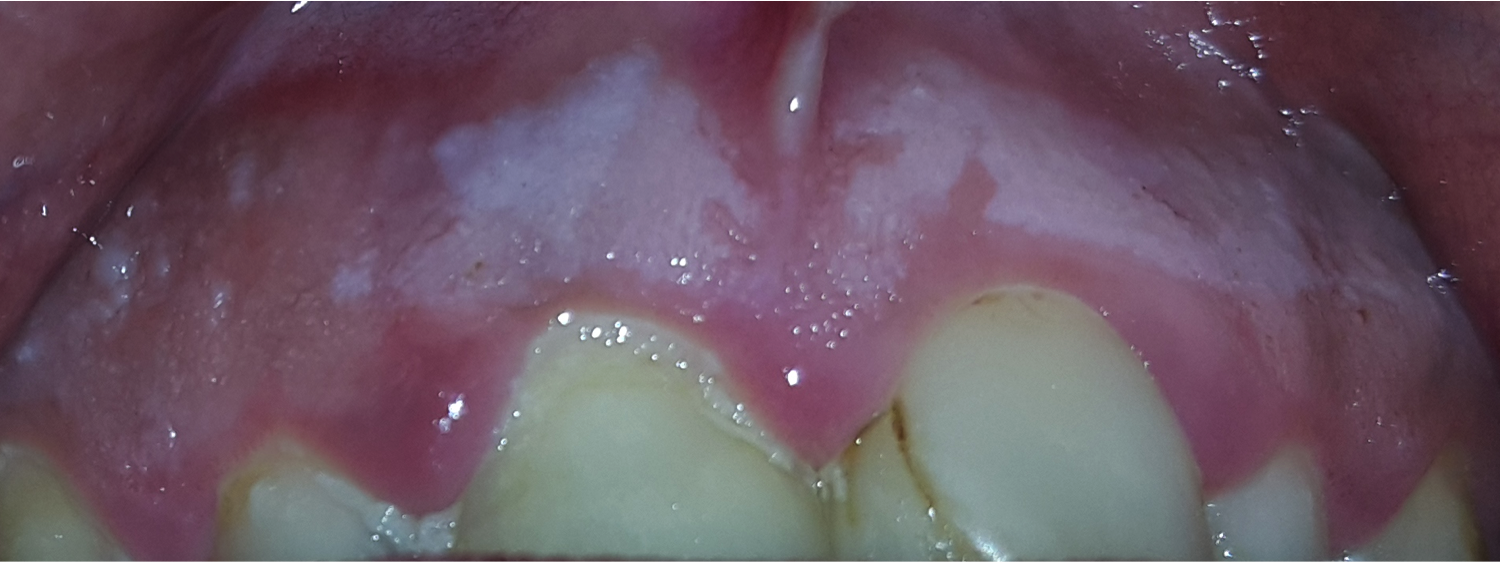
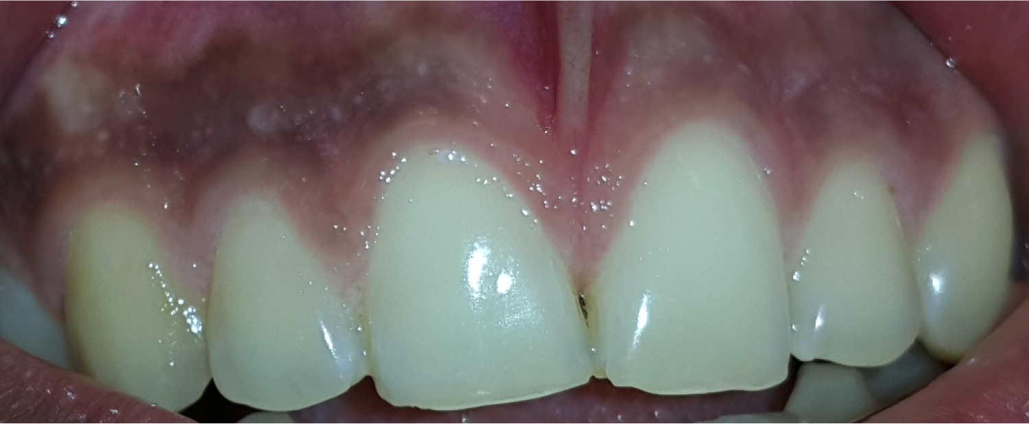
Methods: Two dermatologists described the semiological findings of photographs of the oral cavity of a total of 116 patients (96 patients with Lupus and 20 patients with DM). All patients with SLE fulfilled at least 4 ACR classification criteria. We analyzed 4 areas: gums, soft palate, hard palate and buccal mucosa. For each of these areas the evaluators described the presence of the following clinical findings: red macules, red dots, telangiectasias, pigmented light brown macules, pigmented dark brown macules, discoid lesions, whitish streaks, white patches, mucosal folds, fine cobblestone and thick cobblestone (Fig. 1, 2, 3). We tried to find mucosal clinical features that differentiate patients with strictly CL with those with CL and SLE, and with those with strictly SLE. Also we compared oral features of patients with SLE (with or without skin involvement) of those with DM, since sometimes these 2 clinical entities are very similar. Patients were divided into 3 different groups: SLE with and without cutaneous involvement; CL with and without systemic manifestations and subjects with DM and SLE with or without cutaneous manifestations. X2 or Fisher exact test was used. A p value < 0.05 was considered significant
Results: Of all 84 patients with CL (with or without SLE), discoid mucous patches were only seen in patients with CL with SLE (15.4% vs 0%, p=0.008). Light and dark brown macules on gums were more frequent in patients with CL without SLE (48.3% vs 23.1% p=0.033 and 27.6% vs 7.7% p=0.033, respectively). Of all 38 SLE patients (with or without CL), red spots on hard palate and thick cobblestone on gums were more common in patients with SLE with CL (69.2% vs 33.3% p=0.037 and 53.8% vs 16.7 % p=0.031, respectively). White mucous streaks were more frequent in patients with SLE without CL (41.7% vs 3.8%, p=0.008). When comparing 38 SLE patients and 20 patients with DM, the mucosal folds on hard palate were only in SLE patients (15.8% vs 0%, p=0.084)
Conclusion: There are signs in the oral cavity that help us to differentiate CL from SLE, and SLE from DM  Fig 1: white patch
  Fig 2: discoid lesion
  Fig 3: fine cobblestone
To cite this abstract in AMA style:
Vera-Kellet C Sr., del Barrio-Díaz P Sr., Manríquez-Moreno J Sr., Reyes-Vivanco C Sr.. The Use of Clinical Mucosal Manifestations to Differentiate Patients with Lupus and Dermatomyositis: Transversal, Retrospective and Analytical Study of 116 Patients [abstract]. Arthritis Rheumatol. 2016; 68 (suppl 10). https://acrabstracts.org/abstract/the-use-of-clinical-mucosal-manifestations-to-differentiate-patients-with-lupus-and-dermatomyositis-transversal-retrospective-and-analytical-study-of-116-patients/. Accessed .« Back to 2016 ACR/ARHP Annual Meeting
ACR Meeting Abstracts - https://acrabstracts.org/abstract/the-use-of-clinical-mucosal-manifestations-to-differentiate-patients-with-lupus-and-dermatomyositis-transversal-retrospective-and-analytical-study-of-116-patients/
