ACR Meeting Abstracts

ACR Meeting Abstracts

  • Meetings
    • ACR Convergence 2024
    • ACR Convergence 2023
    • 2023 ACR/ARP PRSYM
    • ACR Convergence 2022
    • ACR Convergence 2021
    • ACR Convergence 2020
    • 2020 ACR/ARP PRSYM
    • 2019 ACR/ARP Annual Meeting
    • 2018-2009 Meetings
    • Download Abstracts
  • Keyword Index
  • Advanced Search
  • Your Favorites
    • Favorites
    • Login
    • View and print all favorites
    • Clear all your favorites
  • ACR Meetings

Abstract Number: 862

Utility of 18f FDG Positron Emission Tomography (PET) As a Diagnostic Test and Marker of Disease Activity in Large Vessel Vasculitis

Sara Alehashemi1, Mark Ahlman2, Ali Cahid Civelek2, Elaine Novakovich3, Ashkan Malayeri2, Peter A. Merkel4, Thomas Cupps5, David Bluemke2 and Peter C. Grayson3, 1Rheumatology, National Institute of Arthritis and Musculoskeletal and Skin Diseases, National Institutes of Health, Bethesda, MD, 2Radiology and Imaging Sciences, National Institutes of Health, Bethesda, MD, 3National Institute of Arthritis and Musculoskeletal and Skin Diseases, National Institutes of Health, Bethesda, MD, 4Division of Rheumatology, Univ of Pennsylvania; Perelman School of Med, Philadelphia, PA, 5Rheumatology, Georgetown University, Bethesda, MD

Meeting: 2016 ACR/ARHP Annual Meeting

Date of first publication: September 28, 2016

Keywords: giant cell arteritis, large vessel vasculitis, positron emission tomography (PET) and takayasu arteritis

  • Tweet
  • Email
  • Print
Session Information

Date: Sunday, November 13, 2016

Title: Vasculitis - Poster I: Large Vessel Vasculitis and Polymyalgia Rheumatica

Session Type: ACR Poster Session A

Session Time: 9:00AM-11:00AM

Background/Purpose: FDG-PET may have a role in diagnosing and monitoring disease activity in large vessel vasculitis (LVV).  Studies that assess the diagnostic accuracy of PET in LVV often use patients with cancer as a comparator group.  There are limited data regarding use of serial FDG-PET imaging to monitor disease activity, and most assessments are performed during periods of clinically apparent disease. This study assesses the diagnostic performance of PET to differentiate LVV from mimics of vasculitis, examines the role of PET as a measure of disease activity, and determines the clinical characteristics associated with abnormal vascular FDG uptake.

Methods: 42 patients with LVV and 50 comparators (15 with systemic inflammation, 35 with atherosclerosis) were recruited in this prospective, observational cohort. Patients with LVV underwent FDG-PET every 6 months.  Clinical and imaging assessments were performed blinded to each other and to levels of acute phase reactants. FDG-PET/CT was performed with 120 min uptake time (256 matrix, 3 mm slice). Active vasculitis on PET was defined by agreement of two nuclear medicine experts and inter-rater agreement was assessed by the kappa statistic. Multivariable regression, adjusted for repeated measures, assessed associations between active vasculitis on PET and clinical characteristics.

Results: 125 FDG-PET scans were performed in 92 patients: giant cell arteritis (n=22; mean age 68.4±9.4 years; disease duration 2.4±2.5 years), Takayasu arteritis (n=20, mean age 34.9±11.2 years; disease duration 13.4±11 years) and comparators (n=50, mean age 53.6±15.3 years).  Inter-rater agreement was high (kappa = 0.84).  In patients with LVV with clinically active disease, the performance characteristics of PET as a diagnostic test were good (sensitivity 90%; specificity 86%); however, 17% of patients with atherosclerosis had PET scan findings indistinguishable from active vasculitis.  26 of 45 PET scans (58%) performed in patients with LVV during apparent clinical remission suggested ongoing active vasculitis.  Clinically apparent active disease and shorter disease duration were significantly associated with an abnormal PET scan.  There was no association between levels of acute phase reactants and PET abnormalities; treatment with immunomodulating medications had a non-statistically significant protective effect (Table).

Conclusion: FDG-PET is useful to distinguish active LVV from mimics of vasculitis, but patients with atherosclerosis can have PET scan findings that closely resemble LVV.  The majority of patients with LVV have abnormal PET scans while in clinical remission.  An inverse association between disease duration and PET abnormalities suggests that scan abnormalities observed during clinical remission are not due to accrued vascular damage.  FDG-PET may be an important modality to detect and monitor subclinical inflammation in LVV. Description: Macintosh HD:Users:alehashemis:Desktop:Screen Shot 2016-06-21 at 9.36.36 AM.png


Disclosure: S. Alehashemi, None; M. Ahlman, None; A. C. Civelek, None; E. Novakovich, None; A. Malayeri, None; P. A. Merkel, None; T. Cupps, None; D. Bluemke, Siemens, 9; P. C. Grayson, None.

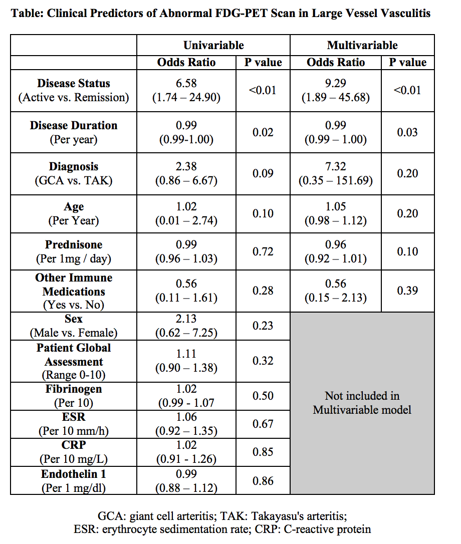
To cite this abstract in AMA style:

Alehashemi S, Ahlman M, Civelek AC, Novakovich E, Malayeri A, Merkel PA, Cupps T, Bluemke D, Grayson PC. Utility of 18f FDG Positron Emission Tomography (PET) As a Diagnostic Test and Marker of Disease Activity in Large Vessel Vasculitis [abstract]. Arthritis Rheumatol. 2016; 68 (suppl 10). https://acrabstracts.org/abstract/utility-of-18f-fdg-positron-emission-tomography-pet-as-a-diagnostic-test-and-marker-of-disease-activity-in-large-vessel-vasculitis/. Accessed .
  • Tweet
  • Email
  • Print

« Back to 2016 ACR/ARHP Annual Meeting

ACR Meeting Abstracts - https://acrabstracts.org/abstract/utility-of-18f-fdg-positron-emission-tomography-pet-as-a-diagnostic-test-and-marker-of-disease-activity-in-large-vessel-vasculitis/

Advanced Search

Your Favorites

You can save and print a list of your favorite abstracts during your browser session by clicking the “Favorite” button at the bottom of any abstract. View your favorites »

All abstracts accepted to ACR Convergence are under media embargo once the ACR has notified presenters of their abstract’s acceptance. They may be presented at other meetings or published as manuscripts after this time but should not be discussed in non-scholarly venues or outlets. The following embargo policies are strictly enforced by the ACR.

Accepted abstracts are made available to the public online in advance of the meeting and are published in a special online supplement of our scientific journal, Arthritis & Rheumatology. Information contained in those abstracts may not be released until the abstracts appear online. In an exception to the media embargo, academic institutions, private organizations, and companies with products whose value may be influenced by information contained in an abstract may issue a press release to coincide with the availability of an ACR abstract on the ACR website. However, the ACR continues to require that information that goes beyond that contained in the abstract (e.g., discussion of the abstract done as part of editorial news coverage) is under media embargo until 10:00 AM ET on November 14, 2024. Journalists with access to embargoed information cannot release articles or editorial news coverage before this time. Editorial news coverage is considered original articles/videos developed by employed journalists to report facts, commentary, and subject matter expert quotes in a narrative form using a variety of sources (e.g., research, announcements, press releases, events, etc.).

Violation of this policy may result in the abstract being withdrawn from the meeting and other measures deemed appropriate. Authors are responsible for notifying colleagues, institutions, communications firms, and all other stakeholders related to the development or promotion of the abstract about this policy. If you have questions about the ACR abstract embargo policy, please contact ACR abstracts staff at [email protected].

Wiley

  • Online Journal
  • Privacy Policy
  • Permissions Policies
  • Cookie Preferences

© Copyright 2025 American College of Rheumatology