Session Information
Session Type: Abstract Submissions (ACR)
Background/Purpose: Medial knee osteoarthritis (MKOA) has been shown to be related to malalignment of the knee joint with several radiographs used to quantify the abnormality. Radiographic observations of a lateral tibial shift in subjects with MKOA has led the authors to hypothesize that this finding is more prevalent in MKOA than normal controls and is associated with MKOA measurable gait parameters.
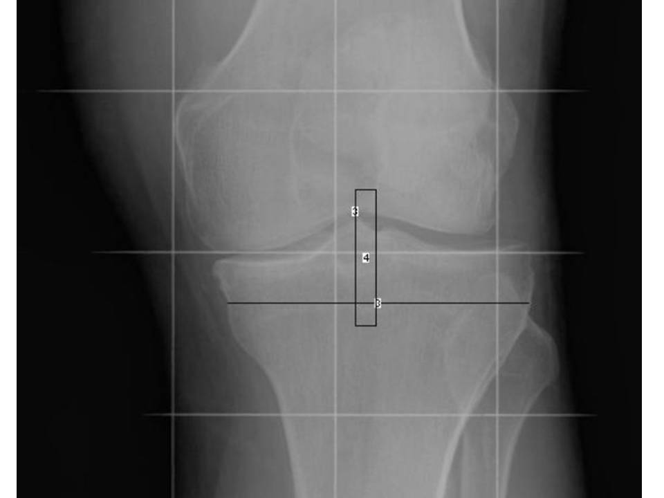
Methods: 90 subjects (69F 21M, Age 60±8, BMI 28.3±4.0) with radiographic and symptomatic medial knee OA (K-L grade 2-3, ambulatory pain >30 mm on a 100 mm VAS) were compared to 24 (18F 6M) age (59±10) and BMI (28.8±8.3) matched controls with no knee pain (K-L grade 0-1). Full limb mechanical axis and AP X-rays of the ankles were obtained. The tibial lateral shift (figure 1), defined as the distance between the center of the intercondylar notch of the femur and midpoint of the tibial plateau, was measured using Image J software (US NIH, Bethesda, MD, http://rsbweb.nih.gov/ij/). Subjects underwent gait analyses using an optoelectronic camera system and multi-component force plate. Comparisons were performed after matching for speed. The peak external knee Adduction Moment (%body weight * height, %BW*Ht) and knee adduction angular impulse was calculated and used as the primary endpoint. Paired t-tests were used to compare group differences. Pearson’s correlations were calculated to analyze the relationship between knee moments and the other radiographic parameters with significance set at p<0.05.
Results: The mean ± S.D. lateral tibio-femoral shift was 5.18±2.45 mm in the MKOA group compared to 1.5±1.22 mm in the control group (p<0.01). Interestingly there was no relationship between the lateral shift and mechanical axis (r=0.11, p=0.23). There was an apparent relationship between the external knee adduction moment and lateral tibial shift in the MKOA group with greater lateral tibial shift related to greater knee moments (r=0.46, p<0.01). There was no relationship between knee adduction moments and lateral tibial shift in the control group (r=0.13, p=0.09). There was a relationship between knee angular impulse to the lateral shift in the MKOA group (r=0.48, p<0.01).
Conclusion: Lateral tibio-femoral shift is greater in MKOA than in normal controls and is related to increased medial knee loads. These findings suggest that the lateral tibio-femoral shift may be a new radiographic marker for MKOA. Further studies are needed to determine the clinical validity of assessing the tibio-femoral shift.
Disclosure:
R. H. Lidtke,
None;
B. Goker,
None;
A. Tufan,
None;
L. E. Thorp,
None;
J. A. Block,
None.
« Back to 2012 ACR/ARHP Annual Meeting
ACR Meeting Abstracts - https://acrabstracts.org/abstract/lateral-tibio-femoral-shift-related-to-medial-knee-osteoarthritis/
