Session Information
Date: Monday, November 9, 2015
Title: Miscellaneous Rheumatic and Inflammatory Diseases Poster Session II
Session Type: ACR Poster Session B
Session Time: 9:00AM-11:00AM
Background/Purpose:
IgG4-related disease (IgG4-RD) is
an immune-mediated fibro-inflammatory condition that often leads to tumefactive lesions.
We describe a common but under-recognized radiologic finding in this
disease: the occurrence of paravertebral masses that may mimic a number of
other inflammatory, infectious, and malignant disorders.
Methods:
The medical records of 143 subjects
with IgG4-RD diagnosed by strict clinicopathologic
correlation were examined to identify those with relevant imaging (n = 59). The available computed
tomography (CT), 18F-fluorodeoxyglucose (18F-FDG) positron emission tomography
(PET), and magnetic resonance imaging (MRI) studies were reviewed by thoracic
radiologists. All images
were reviewed and interpreted by board-certified radiologists. Demographic, clinical, and serological
features were collected from the medical record.
Results:
Of 59 patients with relevant
imaging, 8 (14%) had a paravertebral mass.
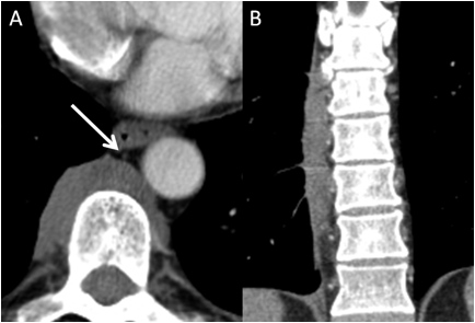
These lesions were typically detected on the right side, along the lower
thoracic spine, and spanned multiple vertebral bodies (Figure 1). The lesions demonstrated
contrast enhancement on CT and 18F-FDG avidity on PET.
All patients not on treatment had
elevated serum IgG4 concentrations.
Multi-organ IgG4-RD was present in seven patients (88%). One of the four patients with imaging
studies before and after treatment demonstrated resolution of the lesions.
Conclusion:
Paravertebral masses are often
detected incidentally on pulmonary imaging in IgG4-RD. Our series of eight cases is, to our
knowledge, the largest series of paravertebral masses associated with IgG4-RD
reported in the literature. All of
our cases had biopsy-proven IgG4-RD. Characteristic features of these lesions include
their right-sided location, the extension across multiple vertebral bodies, and
their common association with other manifestations of IgG4-RD.
Figure
1. (a) Axial contrast enhanced CT in a 59 year old man with IgG4-RD. The paravertebral mass is centered
predominantly on the right side of the thoracic spine. The thoracic duct
(arrow) can be identified anterior to, and separate from, the paravertebral
mass. (b) Follow up CT with coronal
reformats demonstrates the typical craniocaudal
extent, spanning multiple vertebral bodies.
To cite this abstract in AMA style:
Wallace Z, Lim S, Stone JH, McInnis M, Sharma A. Thoracic Paravertebral Masses and IgG4-Related Disease: Report of 8 Cases and Review of the Literature [abstract]. Arthritis Rheumatol. 2015; 67 (suppl 10). https://acrabstracts.org/abstract/thoracic-paravertebral-masses-and-igg4-related-disease-report-of-8-cases-and-review-of-the-literature/. Accessed .« Back to 2015 ACR/ARHP Annual Meeting
ACR Meeting Abstracts - https://acrabstracts.org/abstract/thoracic-paravertebral-masses-and-igg4-related-disease-report-of-8-cases-and-review-of-the-literature/

